
RIN-m5f
Cat.No.: CSC-C9124W
Species: Rattus norvegicus (Rat)
Source: Pancreas
Morphology: epithelial
- Specification
- Background
- Scientific Data
- Q & A
- Customer Review
RIN‑m5F cells are rat pancreatic β‑cell line originally isolated from insulinoma of rat islet of Langerhans. RIN‑m5F Cells exhibit many functional properties of primary pancreatic β-cells including synthesis, storage, and glucose-induced release of insulin as well as responsiveness to other insulin secretagogues. The insulin secreting nature of RIN‑m5F cells make them a popular model for studying pancreatic β-cell physiology in vitro including glucose stimulated insulin secretion, diabetes and mechanisms affecting pancreatic β‑cell function.
RIN‑m5F cells exhibit an epithelial like growth morphology as adherent clusters of cells. RIN-m5F cells also display key β-cell markers, including insulin, Pdx1, and GLUT2, and their function involves insulin release alterations triggered by glucose, amino acids, and drugs. Additionally, these cells can be used to study β-cell stress, oxidative stress and apoptosis.
RIN‑m5F cells have been utilized as models for studying type 1 diabetes and type 2 diabetes including pancreatic β-cell dysfunction, autoimmune-mediated β-cell destruction, drug screening and pancreatic β-cell protective mechanisms. Scientists also utilize RIN-m5F cells to investigate insulin production, ion channel activity, and intracellular signaling pathways.
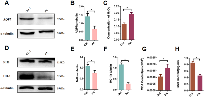
Under High-Lipid Condition, the Expression of AQP7 in RIN-m5f Islet β-Cells was Decreased, Oxidative Stress was Activated, and Ferroptosis Occurred
Aquaporin-7 (AQP7) has been shown to be necessary for pancreatic islet β-cell survival. To study the effects of high lipid conditions on AQP7 expression and downstream effects, Luan et al. utilized RIN-m5f islet β-cells treated with high levels of palmitic acid.
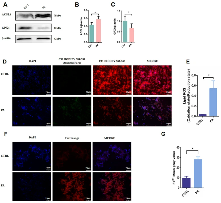
After treatment with palmitic acid AQP7 protein expression was greatly decreased compared to the control group (Fig. 1A-C). High lipid conditions led to an increase in intracellular H₂O₂ (Fig. 1A-C). The cells treated with high lipid conditions showed decreased HO-1 and Nrf2 expression, oxidative stress markers MDA was increased, and cells had lower amounts of GSH (Fig. 1D-H). When assessing ferroptosis during high lipid conditions there was an increase in expression of acyl-CoA synthetase (ACSL4) and a decrease in glutathione peroxidase 4 (GPX4) (Fig. 2A-C). There was also an increase in lipid peroxidation and intracellular Fe²⁺ after high lipid treatment (Fig. 2D-G).
Ask a Question
Write your own review
Description: Established from the pituitary tumor of an ovarectomized F344 rat treated with estrogen for 3 months; described as showing markedly reduced levels of TGF-β1 and TGF-β type II receptor mRNA; ...

Description: This cell line was derived from a 7-month-old female Wistar-Furth rat. GH3 cells produce growth hormone faster than the GH1 cell line and also produce prolactin. Hydrocortisone stimulates growth ...
Description: medullary thyroid carcinoma, neurotensin and calcitonin-producing, (WAG/RIJ rat)

Description: Ethyl nitrosourea induced tumour of the spinal cord and roots from a Wistar-Furth rat. In cell culture the cells grow to a high density and form a sheet of cells with many processes. TR33B is the ...
- Adipose Tissue-Derived Stem Cells
- Human Neurons
- Mouse Probe
- Whole Chromosome Painting Probes
- Hepatic Cells
- Renal Cells
- In Vitro ADME Kits
- Tissue Microarray
- Tissue Blocks
- Tissue Sections
- FFPE Cell Pellet
- Probe
- Centromere Probes
- Telomere Probes
- Satellite Enumeration Probes
- Subtelomere Specific Probes
- Bacterial Probes
- ISH/FISH Probes
- Exosome Isolation Kit
- Human Adult Stem Cells
- Mouse Stem Cells
- iPSCs
- Mouse Embryonic Stem Cells
- iPSC Differentiation Kits
- Mesenchymal Stem Cells
- Immortalized Human Cells
- Immortalized Murine Cells
- Cell Immortalization Kit
- Adipose Cells
- Cardiac Cells
- Dermal Cells
- Epidermal Cells
- Peripheral Blood Mononuclear Cells
- Umbilical Cord Cells
- Monkey Primary Cells
- Mouse Primary Cells
- Breast Tumor Cells
- Colorectal Tumor Cells
- Esophageal Tumor Cells
- Lung Tumor Cells
- Leukemia/Lymphoma/Myeloma Cells
- Ovarian Tumor Cells
- Pancreatic Tumor Cells
- Mouse Tumor Cells