
JL-1
Cat.No.: CSC-C2542
Species: Homo sapiens (Human)
Source: Lung
Morphology: adherent, epithelial-like cells growing in monolayers
Culture Properties: monolayer
- Specification
- Background
- Scientific Data
- Q & A
- Customer Review
Immunology: cytokeratin +, cytokeratin-7 +, cytokeratin-8 +, cytokeratin-17 -, cytokeratin-18 +, cytokeratin-19 +, desmin -, endothel -, GFAP -, neurofilament -, vimentin +
Viruses: PCR: EBV -,
JL-1 is a human-derived continuous cell line originally established from malignant mesothelioma-associated tissue. It is a tumorigenic, adherent cell model that exhibits mixed epithelial- and spindle-like morphology, reflecting the heterogeneity commonly observed in mesothelial-derived malignancies. JL-1 cells display robust proliferative capacity and are widely utilized as an in vitro system for studying the biological behavior of aggressive mesothelioma and related tumor types.
At the molecular level, JL-1 cells retain key characteristics of malignant mesothelial cells and are frequently used in research focusing on cytoskeletal organization, cell adhesion, migration, and invasion. Their stable growth properties and reproducible phenotypic features make them suitable for mechanistic studies investigating tumor progression, epithelial-mesenchymal transition (EMT)-like processes, and tumor microenvironment interactions. In addition, JL-1 cells have been applied in transcriptomic and functional genomic studies to identify dysregulated signaling pathways involved in tumorigenesis.
JL-1 is also used as a model for anticancer drug screening and therapeutic response evaluation, particularly in studies targeting cell motility and survival pathways. Due to its consistent performance in vitro and relevance to mesothelioma biology, JL-1 serves as a valuable experimental platform for both basic research and translational oncology applications, supporting the development of novel therapeutic strategies against highly invasive thoracic tumors.
Pirfenidone Inhibits Mesothelioma Cell Proliferation and Migration
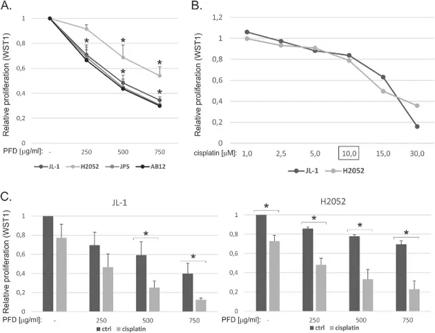
Malignant mesothelioma is an aggressive cancer characterized by a desmoplastic stroma and chemoresistance. Li's team evaluated the anti-tumor potential of pirfenidone, an anti-fibrotic agent. Pirfenidone significantly reduced the proliferation of human (JL-1, H2052, JP5) and mouse (AB12) mesothelioma cell lines after 48 hours (Fig. 1A). A concentration of 750 μg/ml reduced H2052 cell growth by 50%, while other cell lines showed 70% inhibition. Combining pirfenidone with 10 μM cisplatin-the standard chemotherapeutic backbone-resulted in at least an additive reduction in proliferation for JL-1 and H2052 cells (Fig. 1B, C); however, no synergistic effect was observed when combining pirfenidone with pemetrexed.
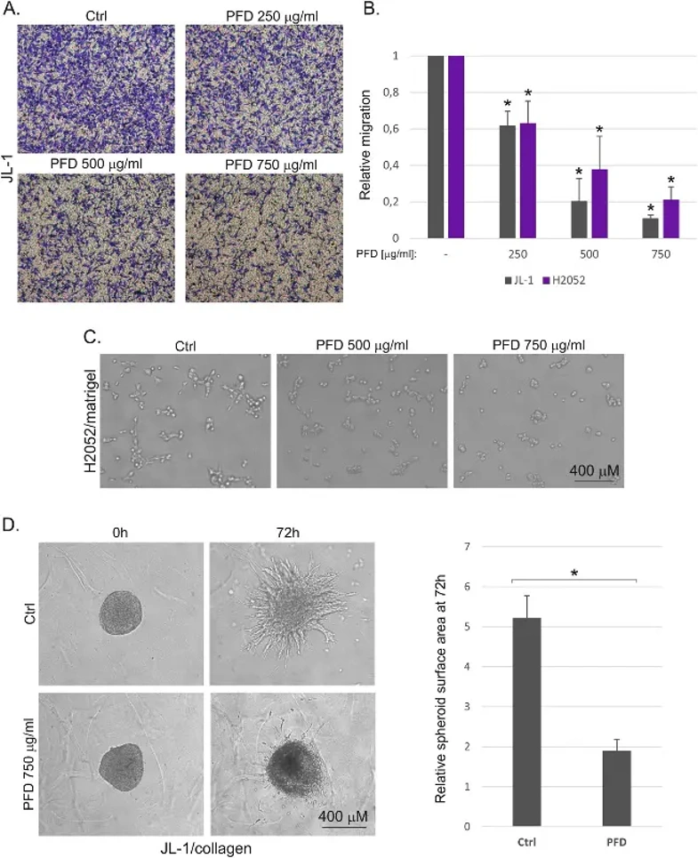
Beyond proliferation, pirfenidone inhibited cell motility. Transwell assays demonstrated a concentration-dependent reduction in JL-1 and H2052 cell migration and invasion through collagen I-coated inserts (Fig. 2A, B). Furthermore, pirfenidone suppressed the invasive sprouting of H2052 cells in 3D Matrigel (Fig. 2C) and reduced the invasive growth of JL-1 cells in a 3D collagen matrix (Fig. 2D). These findings identify pirfenidone as a novel inhibitor of both proliferation and migration in malignant mesothelioma.
Ask a Question
Write your own review
- You May Also Need
Description: Species: human, Caucasian male adult; Tissue: skin; Tumor: melanoma
Description: Species: human male 67 years old; Tissue: pleural effusion; Tumor: mesothelioma
Description: Established from a pleural biopsy of a 62-year-old man with malignant mesothelioma
Description: Species: human, Caucasian male adult; Tissue: skin; Tumor: melanoma
Description: Established in 2003 from the untreated malignant mesothelioma of the pleura of a 78-year-old man with asbestos cancer
Description: established from the pleural effusion of a 71- year- old Caucasian man with pleural epithelioid mesothelioma
- Adipose Tissue-Derived Stem Cells
- Human Neurons
- Mouse Probe
- Whole Chromosome Painting Probes
- Hepatic Cells
- Renal Cells
- In Vitro ADME Kits
- Tissue Microarray
- Tissue Blocks
- Tissue Sections
- FFPE Cell Pellet
- Probe
- Centromere Probes
- Telomere Probes
- Satellite Enumeration Probes
- Subtelomere Specific Probes
- Bacterial Probes
- ISH/FISH Probes
- Exosome Isolation Kit
- Human Adult Stem Cells
- Mouse Stem Cells
- iPSCs
- Mouse Embryonic Stem Cells
- iPSC Differentiation Kits
- Mesenchymal Stem Cells
- Immortalized Human Cells
- Immortalized Murine Cells
- Cell Immortalization Kit
- Adipose Cells
- Cardiac Cells
- Dermal Cells
- Epidermal Cells
- Peripheral Blood Mononuclear Cells
- Umbilical Cord Cells
- Monkey Primary Cells
- Mouse Primary Cells
- Breast Tumor Cells
- Colorectal Tumor Cells
- Esophageal Tumor Cells
- Lung Tumor Cells
- Leukemia/Lymphoma/Myeloma Cells
- Ovarian Tumor Cells
- Pancreatic Tumor Cells
- Mouse Tumor Cells