
IST-MES2
Cat.No.: CSC-6274W
Species: Homo sapiens (Human)
Source: Pleural Effusion
Morphology: grown as monolayer, epithelial-like
Culture Properties: Adherent
- Specification
- Background
- Scientific Data
- Q & A
- Customer Review
Viruses: PCR: HBV -, HCV -, HHV-8 -, HIV-1 -, HIV-2 -, HTLV-I/II -, MLV -, SMRV -
IST-MES2 is a continuously growing human malignant pleural mesothelioma (MPM) cell line established in 1997 from the pleural effusion of a 71-year-old Caucasian male patient with histologically confirmed pleural epithelioid mesothelioma. IST-MES2 exhibits an adherent, epithelial-like morphology with spindle-shaped cells that form a characteristic cobblestone-like pattern at confluence. Karyotypically, the cell line is hypodiploid and displays complex chromosomal alterations, including losses in chromosome arms 9p and 22q, which are commonly associated with mesothelioma pathogenesis.
IST-MES2 expresses canonical mesothelial markers including calretinin, WT-1, and cytokeratin, while lacking EGFR amplification. Notably, the cells also express stemness-associated markers such as CD24, ABCG2, and OCT4, rendering them valuable for investigating mechanisms of drug resistance and metastatic progression in MPM. The cell line constitutively produces IL-6 and TGF-β2, and exhibits positive immunoreactivity for CAM5.2 and HBME-1. IST-MES2 has a doubling time ranging from 37 to 87 hours and reaches exponential growth within 4-7 days post-plating. Importantly, IST-MES2 cells are non-tumorigenic in nude mice upon orthotopic pleural injection, distinguishing this epithelioid line from biphasic counterparts such as IST-MES3 and MM-B1, which readily form pleural tumors.
IST-MES2 serves as a robust in vitro model for studying MPM biology, asbestos-related oncogenesis, and therapeutic resistance. The line has been extensively utilized in preclinical screening of chemotherapeutics, targeted agents, and immunotherapies. Furthermore, the availability of multiple CRISPR-engineered knockout derivatives-targeting genes involved in O-GlcNAcylation (OGT), N-glycan branching (MGAT4C), core fucosylation (FUT8), O-glycosylation (C1GALT1), and glycosaminoglycan biosynthesis (XYLT2)-positions IST-MES2 as a versatile platform for dissecting the role of glycosylation and post-translational modifications in mesothelioma pathogenesis.
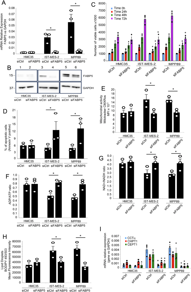
FABP5 Silencing Affects Mesothelioma Cell Dynamics and Metabolism
Pleural mesothelioma (PM) poses a significant challenge in oncology due to its intricate molecular and metabolic landscape, chronic inflammation, and heightened oxidative stress, which contribute to its notorious resilience and clinical complexities. Given the pivotal role attributed to FABP5 and envisioning its potential as a therapeutic target in the context of innovative strategies against mesothelioma, we investigated the consequences of FABP5 silencing on the proliferation, apoptosis, and metabolism of both mesothelial and mesothelioma cells.
Inducing FABP5 silencing, using a pool of siRNA against FABP5 or a scrambled siRNA, as control (Fig. 1A), we observed a notable reduction in the proliferation rates of IstMes2 and MPP89, while the proliferation of normal mesothelial cells HMC35 remained unaffected (Fig. 1B). Remarkably, FABP5 silencing induced a significant increase in apoptosis, as evidenced by Annexin-V assay, in both mesothelioma cell lines IstMes2 and MPP89, while apoptosis levels in normal mesothelial cells remained unaltered (Fig. 1C). Further, we found FABP5 silencing triggered notable metabolic shifts in mesothelioma cells. Specifically, we observed a decrease in mitochondrial activity (Fig. 1D), accompanied by an increase in the ADP/ATP ratio (Fig. 1E) and the NAD/NADH ratio (Fig. 1F). In contrast, normal mesothelial cells exhibited no discernible changes in these metabolic parameters. Furthermore, FABP5 silencing led to a reduction in the abundance of lipid droplets (Fig. 1G) and a concurrent decrease in the mRNA expression levels of key enzymes involved in phospholipid biosynthesis CCTα, CHPT1, and CEPT1 (Fig. 1H). These findings underscore the potential therapeutic efficacy of targeting FABP5, revealing its intricate involvement in mesothelioma cell dynamics and metabolic pathways.
Ask a Question
Write your own review

Description: Species: human, Caucasian male adult; Tissue: skin; Tumor: melanoma
Description: Species: human male 67 years old; Tissue: pleural effusion; Tumor: mesothelioma

Description: Established from a pleural biopsy of a 62-year-old man with malignant mesothelioma

Description: Species: human, Caucasian male adult; Tissue: skin; Tumor: melanoma

Description: Established in 2003 from the untreated malignant mesothelioma of the pleura of a 78-year-old man with asbestos cancer
Description: Species: human, Caucasian male adult; Tissue: skin; Tumor: melanoma
- Adipose Tissue-Derived Stem Cells
- Human Neurons
- Mouse Probe
- Whole Chromosome Painting Probes
- Hepatic Cells
- Renal Cells
- In Vitro ADME Kits
- Tissue Microarray
- Tissue Blocks
- Tissue Sections
- FFPE Cell Pellet
- Probe
- Centromere Probes
- Telomere Probes
- Satellite Enumeration Probes
- Subtelomere Specific Probes
- Bacterial Probes
- ISH/FISH Probes
- Exosome Isolation Kit
- Human Adult Stem Cells
- Mouse Stem Cells
- iPSCs
- Mouse Embryonic Stem Cells
- iPSC Differentiation Kits
- Mesenchymal Stem Cells
- Immortalized Human Cells
- Immortalized Murine Cells
- Cell Immortalization Kit
- Adipose Cells
- Cardiac Cells
- Dermal Cells
- Epidermal Cells
- Peripheral Blood Mononuclear Cells
- Umbilical Cord Cells
- Monkey Primary Cells
- Mouse Primary Cells
- Breast Tumor Cells
- Colorectal Tumor Cells
- Esophageal Tumor Cells
- Lung Tumor Cells
- Leukemia/Lymphoma/Myeloma Cells
- Ovarian Tumor Cells
- Pancreatic Tumor Cells
- Mouse Tumor Cells