

DM-3
Cat.No.: CSC-C2541
Species: Homo sapiens (Human)
Source: Lung
Morphology: adherent, sarcomatoid cells growing in monolayers
Culture Properties: monolayer
- Specification
- Background
- Scientific Data
- Q & A
- Customer Review
Immunology: cytokeratin -, cytokeratin-7 -, cytokeratin-8 -, cytokeratin-17 -, cytokeratin-18 -, desmin -, endothel -, GFAP -, neurofilament -, vimentin +
Viruses: PCR: EBV -, HBV -, HCV -, HIV -
DM-3 is a human malignant pleural mesothelioma cell line generated from a pleural biopsy of a 62-year-old male patient diagnosed with sarcomatoid mesothelioma. The cell line was initially generated as an in vitro model for exploring the biology aspects of malignant mesothelioma, a very aggressive tumor originating from the mesothelial lining of the pleura. DM-3 cells develop in an adherent pattern and have an elongated spindle-shaped morphology consistent with the sarcomatoid subtype of mesothelioma.
DM-3 has morphological and behavioral characteristics consistent with a more mesenchymal cellular state compared to epithelioid mesothelioma models. Studies with DM-3 have explored cell mobility, cytoskeletal architecture, tumor invasiveness, and treatment responsiveness. DM-3 features a highly elongated fibroblast-like morphology and has been utilized to study the association between cytoskeletal architecture and tumor cell motility in analysis of mesothelioma cell panels. DM-3 has also been used in drug response studies as a model to evaluate susceptibility to anticancer drugs (e.g., proteasome inhibitors, redox-modulating chemicals).
DM-3 has also been included into large-scale cancer cell line collections and molecular profiling efforts, enabling study of mesothelioma-related signaling pathways, genomic changes, and treatment vulnerabilities. These features render DM-3 a suitable experimental model for studies in the biology of sarcomatoid mesothelioma, tumor progression, drug screening and translational oncology.
Heterogeneous Actin Cytoskeleton Organization Across Malignant Mesothelioma Cell Lines
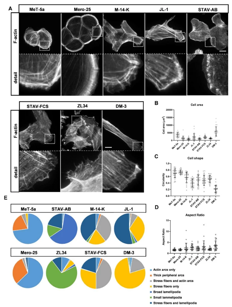
The cytoskeleton is fundamental to tissue homeostasis, controlling cell morphogenesis and migration. Cancer cells show migratory and invasive behaviors that are promoted by dysregulated cytoskeletal dynamics. To study this in MM, Keller et al. analyzed actin filament organization in epithelioid, sarcomatoid and biphasic subtypes of malignant mesothelioma (MM) cell lines.
Cells were seeded on coverslips and after 24 h they were fixed and stained with TRITC-conjugated phalloidin to visualize filamentous actin (F-actin). Epithelioid lines MeT-5A and Mero-25 produced colonies with cortical actin arcs at the periphery, and were tiny and rounded (aspect ratio ~1.6) (Fig. 1A). However, the sarcomatoid line DM-3 showed a fibroblastic morphology with high aspect ratio (3.6) with conspicuous stress fibres throughout the cell (Fig. 1A, C, D). Biphasic lines showed distinct actin organization: JL-1 and STAV-FCS were more elongated (aspect ratios 2.6 and 3.0, respectively) with conspicuous stress fibres whereas STAV-AB had broad lamellipodia at cell borders. Colonies of M-14-K were growing like epithelioid cells while ZL34 had short stress fibres and little condensed membrane ruffles (Fig. 1A). Perinuclear actin caps, short actin filaments capping the nucleus, have been proposed as a structural signature separating epithelial from fibroblast-like cells. Actin caps were extensively established in control BJ fibroblasts, but were not observed in MeT-5A, Mero-25, STAV-AB, and ZL34. For some cells, DM-3, M-14-K, and STAV-FCS had actin caps, while JL-1 retained well-developed caps, similar to fibroblasts.
These results showed the existence of a marked variability in the organization of the actin cytoskeleton in MM cell lines, in agreement with their different morphological and motile features, and suggest the complexity of the cytoskeleton control in the biology of mesothelioma.
Ask a Question
Write your own review
- You May Also Need

Description: Species: human, Caucasian male adult; Tissue: skin; Tumor: melanoma

Description: Species: human male 67 years old; Tissue: pleural effusion; Tumor: mesothelioma

Description: Species: human, Caucasian male adult; Tissue: skin; Tumor: melanoma
Description: Established in 2003 from the untreated malignant mesothelioma of the pleura of a 78-year-old man with asbestos cancer

Description: established from the pleural effusion of a 71- year- old Caucasian man with pleural epithelioid mesothelioma

Description: Species: human, Caucasian male adult; Tissue: skin; Tumor: melanoma
- Adipose Tissue-Derived Stem Cells
- Human Neurons
- Mouse Probe
- Whole Chromosome Painting Probes
- Hepatic Cells
- Renal Cells
- In Vitro ADME Kits
- Tissue Microarray
- Tissue Blocks
- Tissue Sections
- FFPE Cell Pellet
- Probe
- Centromere Probes
- Telomere Probes
- Satellite Enumeration Probes
- Subtelomere Specific Probes
- Bacterial Probes
- ISH/FISH Probes
- Exosome Isolation Kit
- Human Adult Stem Cells
- Mouse Stem Cells
- iPSCs
- Mouse Embryonic Stem Cells
- iPSC Differentiation Kits
- Mesenchymal Stem Cells
- Immortalized Human Cells
- Immortalized Murine Cells
- Cell Immortalization Kit
- Adipose Cells
- Cardiac Cells
- Dermal Cells
- Epidermal Cells
- Peripheral Blood Mononuclear Cells
- Umbilical Cord Cells
- Monkey Primary Cells
- Mouse Primary Cells
- Breast Tumor Cells
- Colorectal Tumor Cells
- Esophageal Tumor Cells
- Lung Tumor Cells
- Leukemia/Lymphoma/Myeloma Cells
- Ovarian Tumor Cells
- Pancreatic Tumor Cells
- Mouse Tumor Cells