
HCEC-12
Cat.No.: CSC-C3456
Species: Homo sapiens (Human)
Source: Eye; Cornea
Morphology: epitheloid cells growing adherently in monolayers
Culture Properties: monolayer
- Specification
- Background
- Scientific Data
- Q & A
- Customer Review
Immunology: cytokeratin +, cytokeratin-7 +, cytokeratin-8 +, cytokeratin-17 +, cytokeratin-18 +, cytokeratin-19 -, desmin -, endothel -, EpCAM -, GFAP-, neurofilament -, vimentin +
Viruses: PCR:
HCEC-12 is a well-characterized, immortalized human corneal endothelial cell line established in 1996. It was derived from the posterior corneal epithelium of a 91-year-old Caucasian woman and immortalized via transformation with the SV40 early region, which encodes the large T-antigen and small t-antigen. The cells exhibit an adherent, epithelioid morphology, growing in monolayers.
The primary advantage of HCEC-12 is its provision of a consistent, renewable source of human corneal endothelial cells (HCEnCs), overcoming the significant limitations of primary cells which have poor proliferative capacity in vivo. HCEC-12 cells retain key physiological characteristics, serving as a valuable in vitro model for studying corneal endothelial biology. For instance, they have been instrumental in elucidating the role of mechanotransduction, demonstrating stiffness-dependent responses that regulate proliferation and phenotype. They also express functional temperature-sensitive TRPV channels, which are essential for corneal endothelial homeostasis. Furthermore, the extracellular matrix (ECM) produced by HCEC-12 can stimulate the growth of primary HCEnCs, highlighting its utility in developing culture systems.
Consequently, HCEC-12 is extensively utilized in research areas including corneal tissue engineering, drug screening, and studies on cell replacement therapy. Its well-documented profile and wide availability make it an indispensable platform for advancing ocular surface and corneal research.
In Vitro Ocular Cytotoxicity of BEE Venom in HCEC-12 Cell Lines
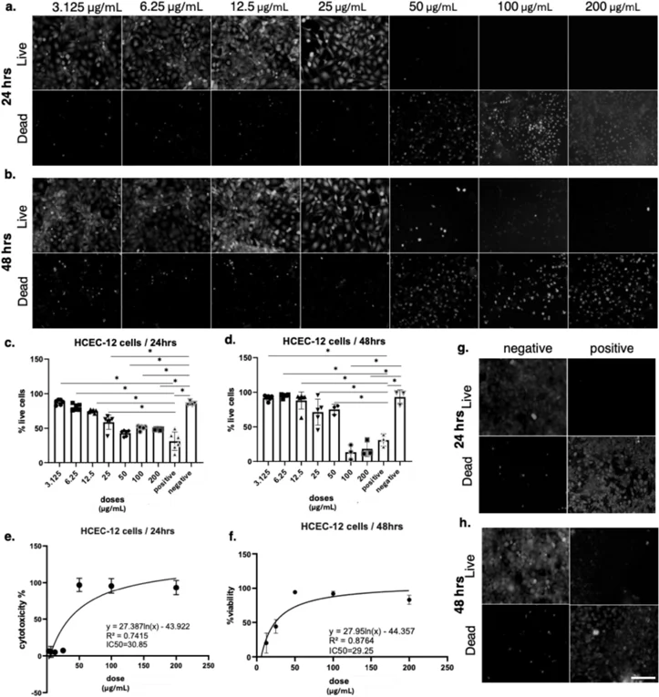
Bee venom (BV) is a complex natural secretion of Apis mellifera with documented anti-inflammatory, antimicrobial, and anticancer properties. Despite its therapeutic potential, safety concerns remain due to its strong cytotoxic effects, particularly in sensitive tissues such as the eye. This study evaluated the cytotoxicity of purified BV in human corneal endothelial cells (HCEC-12) using MTT viability assays and live/dead staining over 24 h and 48 h.
Results showed that at 24 h, cells exposed to concentrations up to 12.5 µg/mL largely maintained viability, whereas 25 µg/mL induced a moderate reduction in live cells. At 50 µg/mL and higher, cell death became evident, with almost complete loss of viability at 100 and 200 µg/mL. After 48 h, the toxic response was stronger, with visible reductions in viability beginning at 12.5 µg/mL, pronounced cell death at 25 µg/mL, and near-total loss of viable cells at concentrations ≥ 100 µg/mL. MTT assay measurements confirmed these findings. Statistical analysis indicated significant differences between treatment groups and untreated controls, confirming the dose-dependent effect.
Ask a Question
Write your own review
- You May Also Need
Description: Established in 2007 from the large retrosternal mass resected before treatment from a 57-year-old Caucasian man with rapidly fatal anaplastic thyroid cancer
Description: established from the uvea tissue of a male patient with uveal melanoma
Description: NTERA-2 was cloned from cell line TERA-2 which was derived from a metastatic teratocarcinoma of a 22-year-old Caucasian male; cell line also known as NT-2
Description: Established from the bone marrow of a 27-year-old woman with B-cell non-Hodgkin lymphoma (B-NHL) (diffuse large cell lymphoma, DLCL, stage 4B, at relapse) in 1987
- Adipose Tissue-Derived Stem Cells
- Human Neurons
- Mouse Probe
- Whole Chromosome Painting Probes
- Hepatic Cells
- Renal Cells
- In Vitro ADME Kits
- Tissue Microarray
- Tissue Blocks
- Tissue Sections
- FFPE Cell Pellet
- Probe
- Centromere Probes
- Telomere Probes
- Satellite Enumeration Probes
- Subtelomere Specific Probes
- Bacterial Probes
- ISH/FISH Probes
- Exosome Isolation Kit
- Human Adult Stem Cells
- Mouse Stem Cells
- iPSCs
- Mouse Embryonic Stem Cells
- iPSC Differentiation Kits
- Mesenchymal Stem Cells
- Immortalized Human Cells
- Immortalized Murine Cells
- Cell Immortalization Kit
- Adipose Cells
- Cardiac Cells
- Dermal Cells
- Epidermal Cells
- Peripheral Blood Mononuclear Cells
- Umbilical Cord Cells
- Monkey Primary Cells
- Mouse Primary Cells
- Breast Tumor Cells
- Colorectal Tumor Cells
- Esophageal Tumor Cells
- Lung Tumor Cells
- Leukemia/Lymphoma/Myeloma Cells
- Ovarian Tumor Cells
- Pancreatic Tumor Cells
- Mouse Tumor Cells