
NTERA-2
Cat.No.: CSC-C0535
Species: Homo sapiens (Human)
Source: Lung Metastasis
Morphology: adherent epitheloid cells carrying strong granula growing adherent in monolayer
Culture Properties: monolayer
- Specification
- Background
- Scientific Data
- Q & A
- Customer Review
Immunology: cytokeratin +, cytokeratin-7 -, cytokeratin-8 +, cytokeratin-17 -, cytokeratin-18 -, cytokeratin-19 +, desmin -, endothel -, EpCAM +, GFAP -, neurofilament -, vimentin -
Viruses: PCR:
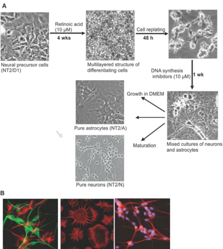
NTERA-2 (NT2) is a pluripotent human embryonal carcinoma (EC) cell line. It was originally isolated from a malignant teratocarcinoma that had grown in the lung of a male patient. As an EC line, it is a well-characterized in vitro model of human embryonic stem cells and has the ability to differentiate into cells of all three germ layers.
NTERA-2 cells are functionally pluripotent. They express the markers SSEA-3, SSEA-4, TRA-1-60, and TRA-1-81. The most relevant functional feature of NTERA-2 cells, however, is their ability to differentiate in response to retinoic acid (RA) treatment. Upon RA treatment, a mixed population of neurons and glial cells is generated with a postmitotic, neuron-like cell becoming the majority of the cells present. NT2 neurons generated in this way express typical neuronal markers, including neurotransmitters, neurofilaments, and functional NMDA and GABA receptors and thus serve as a primary model system for the study of neuronal development and function.
As a result, serves as an essential tool for studying both neuroscientific topics and developmental biology. The line is most often used to study neuronal differentiation, neurotoxicity, neurodegeneration, and neural development. In addition, since they can be infected with JC virus, they are also used as a model system for progressive multifocal leukoencephalopathy (PML). The reliability of the line and the robustness of the differentiation protocol also make this line useful for high-throughput drug screens and for studies on mechanisms of action of neuroactive compounds.
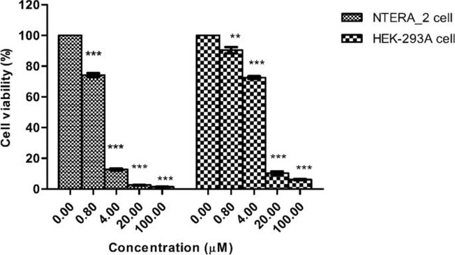
Glyasperin A from Macaranga indica Presents Promising Capacities Against NTERA-2 Cancer Stem Cells
Glyasperin A is a flavonoid that was isolated from the leaves of Macaranga indica Wight, Euphorbiaceae, a Vietnamese traditional herbal medicine. This compound has been proven to have potential anticancer properties. However, its impact on cancer stem cells has not been identified.
Mai et al. tested the cytotoxicity potential of glyasperin A against the NTERA-2 pluripotent human embryonal carcinoma cell line. As shown in Fig. 1, the cytotoxicity of glyasperin A is dose-dependent. For NTERA-2, glyasperin A at the concentrations of 20 µM and 100 µM strictly inhibited the cell viability compared with the negative control. The NTERA-2 cell viability slightly increased when the concentration of the compound decreased to 4 µM. However, the proportion of cell growth was still lower than 50%. In contrast, the cytotoxicity of GLA on HEK-293A cells was less than that on NTERA-2 at all tested concentrations. Notably, the IC50 value for NTERA-2 cells treated with glyasperin A was 2 ± 0.009 µM, while this value for HEK-293A cells was 6.4 ± 0.09 µM. These findings proposed that the NTERA-2 cell line seemed to be more sensitive to glyasperin A than the HEK-293A cell line.
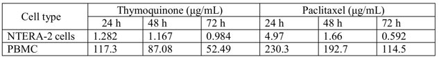
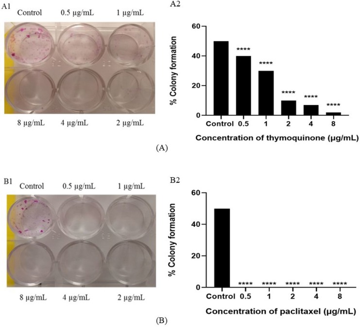
Thymoquinone Affected the Proliferation and Colony Formation Ability of NTERA-2 Cells
Thymoquinone (TQ), the main active constituent of N. sativa, is reported to have anticancer activity. However, the anticancer effects of TQ on the cancer stem cell-like cells are not known. In the present study, the anti-proliferative effect and apoptosis-inducing capacity of TQ in human embryonal carcinoma cells (NTERA-2) and peripheral blood mononuclear cells (PBMCs) were assessed. TQ significantly inhibited the proliferation of NTERA-2 cells (IC50: 1.282, 1.167 and 0.984 µg/mL at 24, 48 and 72 hours respectively) (Fiig. 2). Paclitaxel used as positive control exhibited IC50 values of 4.97, 1.66 and 0.592 µg/mL at 24, 48 and 72 hours, respectively. Paclitaxel and TQ were less toxic in PBMCs (Fig. 2). TQ significantly decreased the colony formation of NTERA-2 in dose-dependent manner in comparison with the controls (Fig. 3A), where as cells treated with Paclitaxel did not form any colonies (0%) (Fig. 3B). These TQ-treated single cells completely lost the ability of division and proliferation, which is a hallmark of potent inhibition of NTERA-2 to form colonies.
Ask a Question
Write your own review
- You May Also Need
Description: Established in 2007 from the large retrosternal mass resected before treatment from a 57-year-old Caucasian man with rapidly fatal anaplastic thyroid cancer

Description: established from the uvea tissue of a male patient with uveal melanoma
Description: Established from the bone marrow of a 27-year-old woman with B-cell non-Hodgkin lymphoma (B-NHL) (diffuse large cell lymphoma, DLCL, stage 4B, at relapse) in 1987

Description: An Epstein-Barr virus (EBV) transformed lymphoblastoid cell line with Human Leukocyte Antigen (HLA)-typing data available.
- Adipose Tissue-Derived Stem Cells
- Human Neurons
- Mouse Probe
- Whole Chromosome Painting Probes
- Hepatic Cells
- Renal Cells
- In Vitro ADME Kits
- Tissue Microarray
- Tissue Blocks
- Tissue Sections
- FFPE Cell Pellet
- Probe
- Centromere Probes
- Telomere Probes
- Satellite Enumeration Probes
- Subtelomere Specific Probes
- Bacterial Probes
- ISH/FISH Probes
- Exosome Isolation Kit
- Human Adult Stem Cells
- Mouse Stem Cells
- iPSCs
- Mouse Embryonic Stem Cells
- iPSC Differentiation Kits
- Mesenchymal Stem Cells
- Immortalized Human Cells
- Immortalized Murine Cells
- Cell Immortalization Kit
- Adipose Cells
- Cardiac Cells
- Dermal Cells
- Epidermal Cells
- Peripheral Blood Mononuclear Cells
- Umbilical Cord Cells
- Monkey Primary Cells
- Mouse Primary Cells
- Breast Tumor Cells
- Colorectal Tumor Cells
- Esophageal Tumor Cells
- Lung Tumor Cells
- Leukemia/Lymphoma/Myeloma Cells
- Ovarian Tumor Cells
- Pancreatic Tumor Cells
- Mouse Tumor Cells