Grunt Fin (GF)
Cat.No.: CSC-C9052H
Species: Haemulon sciurus (Bluestriped grunt)
Source: Fin
Morphology: Fibroblast-like
Culture Properties: Adherent
- Specification
- Background
- Scientific Data
- Q & A
- Customer Review
The Grunt Fin (GF) cell line is a spontaneously immortalized fibroblast-like line derived from the caudal/pectoral fin tissue of an adult saltwater Bluestriped Grunt (Haemulon sciurus), first established in 1961. It grows as an adherent monolayer with a modal chromosome number of 47 and is cultured at 20-25°C in EMEM (with elevated NaCl to mimic seawater osmolarity) supplemented with 10-20% FBS. The line is sensitive to plastic type and has relatively low plating efficiency post-thaw.
In the literature, GF cells are a cornerstone model in aquaculture virology and fish disease research. They are one of the preferred cell lines for the isolation and propagation of megalocytiviruses (Red Sea Bream Iridovirus, RSIV) and have been used to produce the formalin-inactivated RSIV vaccine-a commercially significant fish viral vaccine. GF cells are also employed in aquatic ecotoxicology, serving as an in vitro platform for assessing cytotoxicity and genotoxicity of environmental pollutants (heavy metals, pesticides, microplastics) on marine teleost cells. Additionally, they are used in fish cell senescence, DNA repair, and comparative genomics studies, and as a non-mammalian baseline for evaluating the bio-compatibility of biomaterials in marine contexts.
Establishment and Characterization of a Dwarf Gourami Fin (DGF) Cell Line for Iridovirus Propagation
Fish-derived primary cell lines are invaluable for virological studies due to their rapid growth and high viral susceptibility. Jeong et al. established a novel cell line, dwarf gourami fin (DGF), derived from the caudal fin of Trichogaster lalius, and characterized its optimal growth conditions, karyotype, transfection efficiency, and viral susceptibility.
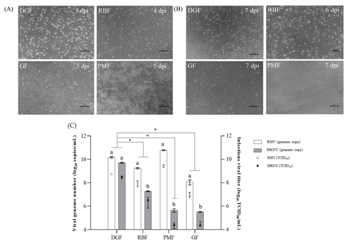
Viral susceptibility assays revealed distinct cytopathic effects (CPE) across cell lines. Following red sea bream iridovirus (RSIV) inoculation, typical megalocytivirus CPE (cell rounding, shrinking, detachment) was observed in DGF, RBF, and GF cells within 7 days, whereas PMF cells exhibited only rounding (Fig. 1A). Infectious spleen and kidney necrosis virus (ISKNV) induced weak CPE in GF cells, but none in PMF cells. Notably, DGF and RBF cells exhibited extensive cell enlargement and rounding by 4 days post-infection (dpi) with ISKNV, progressing to monolayer disruption in RBF cells, though DGF cells retained monolayer integrity through 7 dpi (Fig. 1B).
Quantification of supernatants at 7 dpi revealed distinct viral replication profiles (Fig. 1C). For RSIV, PMF cells yielded the highest titers (5.98 × 10¹⁰ genome copies/mL; 109.44 TCID₅₀/mL), approximately fivefold higher than DGF cells, though infectious titers were not statistically different. Conversely, DGF cells supported the highest ISKNV replication (5.22 × 10⁹ genome copies/mL; 10⁸·⁴⁸ TCID₅₀/mL), which was approximately 100-fold greater than in RBF cells and significantly higher than in GF cells (3.27 × 10⁵ genome copies/mL). These results demonstrate that the DGF cell line is a highly susceptible and efficient model for ISKNV propagation.
Ask a Question
Write your own review
- You May Also Need


Description: Derived from tissue posterior to the anus of normal adult minnows. Reported to be capable of growth over a wide temperature range from 4°C-36°C with maximum growth at 34°C. Support growth of the ...

Description: E11 is a clone of the cell line SSN-1 and is persistantly infected with a C-type retrovirus (SnRV). It is susceptible to piscine nodavirus strains belonging to different genotypes (SJNNV, RGNNV, ...

Description: The fish cell line SSN-1 was initiated from whole fry tissue, Channa (Ophicephalus) striatus, commonly named 'striped snakehead'. SSN-1 spontaneously produce and release endogenous snakehead fish ...

Description: From pooled male/female gonad tissue of yearling rainbow trout (Oncorhynchus mykiss). First cell line to be established from cold-blooded vertebrates. 8-fold difference in growth rate in temperature ...

- Adipose Tissue-Derived Stem Cells
- Human Neurons
- Mouse Probe
- Whole Chromosome Painting Probes
- Hepatic Cells
- Renal Cells
- In Vitro ADME Kits
- Tissue Microarray
- Tissue Blocks
- Tissue Sections
- FFPE Cell Pellet
- Probe
- Centromere Probes
- Telomere Probes
- Satellite Enumeration Probes
- Subtelomere Specific Probes
- Bacterial Probes
- ISH/FISH Probes
- Exosome Isolation Kit
- Human Adult Stem Cells
- Mouse Stem Cells
- iPSCs
- Mouse Embryonic Stem Cells
- iPSC Differentiation Kits
- Mesenchymal Stem Cells
- Immortalized Human Cells
- Immortalized Murine Cells
- Cell Immortalization Kit
- Adipose Cells
- Cardiac Cells
- Dermal Cells
- Epidermal Cells
- Peripheral Blood Mononuclear Cells
- Umbilical Cord Cells
- Monkey Primary Cells
- Mouse Primary Cells
- Breast Tumor Cells
- Colorectal Tumor Cells
- Esophageal Tumor Cells
- Lung Tumor Cells
- Leukemia/Lymphoma/Myeloma Cells
- Ovarian Tumor Cells
- Pancreatic Tumor Cells
- Mouse Tumor Cells