
RTG-2
Cat.No.: CSC-C9056H
Species: Oncorhynchus mykiss (Rainbow trout)
Source: Ovary
Morphology: Fibroblast
Culture Properties: Adherent
- Specification
- Background
- Scientific Data
- Q & A
- Customer Review
RTG-2 is a continuous fibroblast-like cell line originally established from gonadal tissue of the rainbow trout (Oncorhynchus mykiss). It is one of the most commonly used fish cell lines in the fields of aquatic biology, virology and environmental toxicology. RTG-2 cells have well-defined growth characteristics and are highly susceptible to a wide range of fish viruses. The cell line has been widely characterized and is routinely used in fish health and disease research as a standard in vitro model.
Functionally, RTG-2 cells are highly permissive to many fish viruses, including infectious hematopoietic necrosis virus (IHNV), viral hemorrhagic septicemia virus (VHSV) and infectious pancreatic necrosis virus (IPNV), among others. RTG-2 provides a useful platform for virus isolation, propagation and antiviral screening. In addition, the cell line is often used in research studies of fish immunology, host-pathogen interactions and cellular responses to environmental stressors.
RTG-2 is also commonly used in aquatic toxicology and ecotoxicology studies to assess the cytotoxic and genotoxic effects of environmental contaminants, chemicals and pharmaceuticals. Overall, RTG-2 offers a robust, reproducible and biologically relevant in vitro model for research applications in aquaculture, fish virology, environmental safety and regulatory testing.
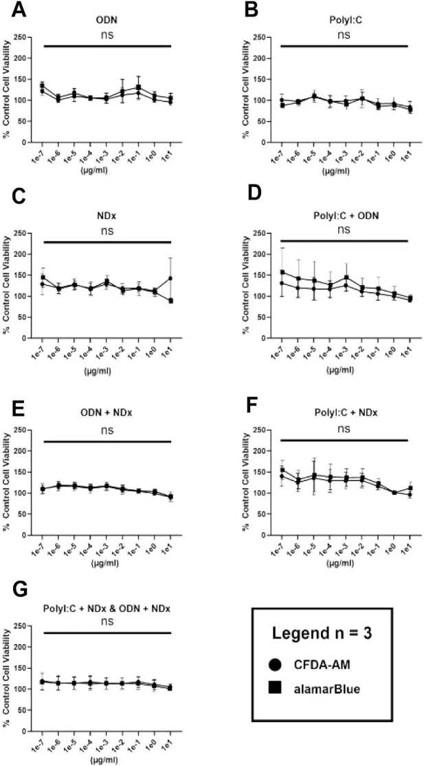
Nucleic Acids Delivered by Cationic Phytoglycogen Nanoparticles Protect RTG-2 Cells Against VHSV-Iva Infection
The innate immune system recognizes pathogen-associated molecular patterns (PAMPs) to defend against infections. Oberhoffner et al. investigated the use of synthetic PAMP analogues to stimulate innate antiviral responses in rainbow trout cells, exploring their potential as prophylactic agents against viral diseases in aquaculture.
The cytotoxicity capacity of each nucleic acid and NDx alone or complexed was measured in vitro. RTG-2 cells were treated with serial dilutions of different nucleic acid and nanoparticle combinations ranging from 0.1 pg to 10 µg/mL (Fig. 1A-G). After 24 hrs, CFDA-AM and alamarBlue, which measure cellular membrane integrity and metabolism respectively, were applied. Neither membrane integrity nor metabolism were significantly reduced by any of the treatment groups at any of the tested concentrations.
Ask a Question
Write your own review
- You May Also Need


Description: Derived from tissue posterior to the anus of normal adult minnows. Reported to be capable of growth over a wide temperature range from 4°C-36°C with maximum growth at 34°C. Support growth of the ...
Description: E11 is a clone of the cell line SSN-1 and is persistantly infected with a C-type retrovirus (SnRV). It is susceptible to piscine nodavirus strains belonging to different genotypes (SJNNV, RGNNV, ...

Description: The fish cell line SSN-1 was initiated from whole fry tissue, Channa (Ophicephalus) striatus, commonly named 'striped snakehead'. SSN-1 spontaneously produce and release endogenous snakehead fish ...


- Adipose Tissue-Derived Stem Cells
- Human Neurons
- Mouse Probe
- Whole Chromosome Painting Probes
- Hepatic Cells
- Renal Cells
- In Vitro ADME Kits
- Tissue Microarray
- Tissue Blocks
- Tissue Sections
- FFPE Cell Pellet
- Probe
- Centromere Probes
- Telomere Probes
- Satellite Enumeration Probes
- Subtelomere Specific Probes
- Bacterial Probes
- ISH/FISH Probes
- Exosome Isolation Kit
- Human Adult Stem Cells
- Mouse Stem Cells
- iPSCs
- Mouse Embryonic Stem Cells
- iPSC Differentiation Kits
- Mesenchymal Stem Cells
- Immortalized Human Cells
- Immortalized Murine Cells
- Cell Immortalization Kit
- Adipose Cells
- Cardiac Cells
- Dermal Cells
- Epidermal Cells
- Peripheral Blood Mononuclear Cells
- Umbilical Cord Cells
- Monkey Primary Cells
- Mouse Primary Cells
- Breast Tumor Cells
- Colorectal Tumor Cells
- Esophageal Tumor Cells
- Lung Tumor Cells
- Leukemia/Lymphoma/Myeloma Cells
- Ovarian Tumor Cells
- Pancreatic Tumor Cells
- Mouse Tumor Cells