
SSN-1
Cat.No.: CSC-C9060H
Species: Channa striata (Snakehead murrel)
Morphology: Fibroblast
Culture Properties: Adherent
- Specification
- Background
- Scientific Data
- Q & A
- Customer Review
SSN-1 is a cell line originally established from fry tissue of striped snakehead (Channa striatus). This cell line is spontaneously immortalized with persistent infection of a type C retrovirus called Snakehead Retrovirus (SnRV). The SSN-1 cell line displays fibroblast morphological properties in cell culture and grows adherently. Researchers use the SSN-1 cell line extensively in laboratories for studying fish viruses. It is very sensitive to many fish viruses such as Betanodavirus (Viral Nervous Necrosis, VNN), Epizootic Ulcerative Syndrome (EUS) virus and Viral Nervous Necrosis.
For example, in virus isolation and identification studies, diseased grouper tissue homogenates were inoculated into SSN-1 cells and obvious cytopathic effects (CPE) were produced in the culture, and NNV-specific bands were identified through RT-PCR methods. In viral replication and pathogenic mechanism studies, transcriptomic and proteomic approaches revealed that NNV infection of SSN-1 cells activated the host interferon system and triggered apoptosis in host cells. The SSN-1 cell line has also been used to screen drugs against NNV. For example, the SSN-1 cell line was used to screen various traditional Chinese medicines with anti-NNV activity, and their activity was determined by CPE variation assessment.
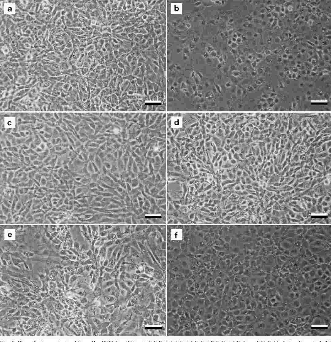 Fig. 1. Six cell clones derived from the SSN-1 cell line (Iwamoto T, Nakai T, et al., 2000).
Correlation between NNV Infectivity and Cytopathic Effect (CPE) Appearance Ratio
Nervous necrosis virus (NNV), a simple spherical RNA virus in the family Nodaviridae, is pathogenic to many fish species. Lee et al. studied the adsorption ratio and infection efficiency of purified NNV on striped snakehead cells (SSN-1) using a 96-well titration system.
The purified NNV suspension (4.0 TCID50/100 µl) was serially diluted twice, and infectivity titration was performed using the 96-well system (Fig. 1). The infectivity titer of the NNV suspension, calculated using a modified Kärber's formula based on the CPE appearance ratio at each dilution, was 4.56 TCID50/100 µl (average of two replicates: 4.46 and 4.66 TCID50/100 µl) (Fig. 1A). The CPE appearance ratio, which is the proportion of CPE-positive wells among all inoculated wells at each dilution, indicates the probability of NNV infection. Plotting these data on a semilogarithmic graph with CPE appearance ratio (%) on the Y axis and NNV infectivity (log10 TCID50/100 µl) on the X axis (Fig. 1B) yielded a regression line (y = 55.7x + 50.6, R2 = 0.9930), showing a close relationship between CPE appearance ratio and NNV infectivity titer. This line indicated that the CPE appearance ratio decreased by 17% each time the NNV infectivity dose was halved. Subsequent NNV infectivity titers were calculated using this regression line with the 96-well system.
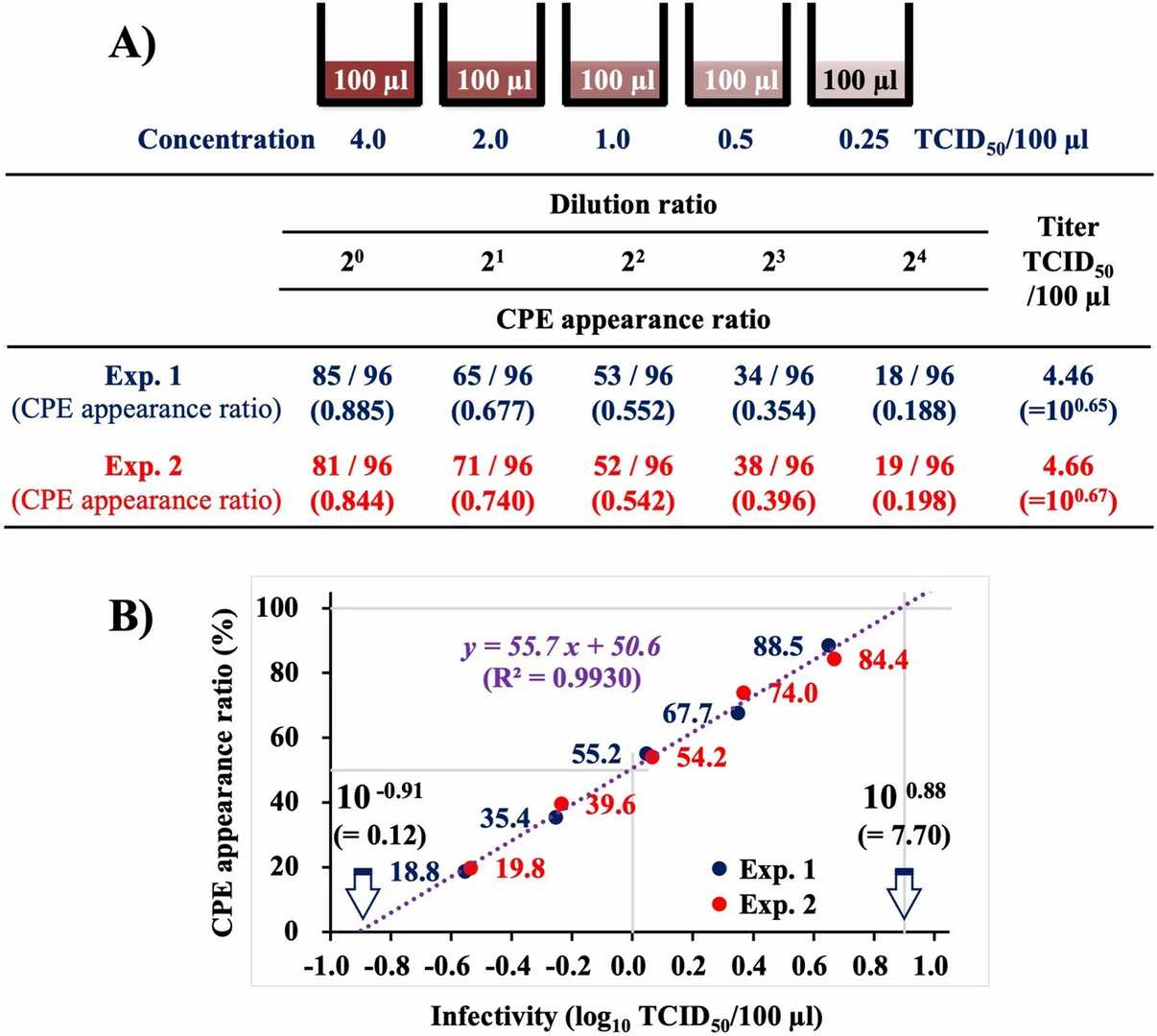 Fig. 1. Correlation of NNV infectivity with CPE appearance ratio demonstrated by titration using the 96-well system (Lee H S, Gye H J, et al., 2023).
Cytopathic Effects in SSN-1 Cells Inoculated with the Filtrate of Naturally Infected Murry Cod Fry Tissue Samples
In December 2019, a severe mass mortality event occurred among cultured Murray cod fry in Foshan city, China, with a cumulative mortality rate reaching 45% within 15 days. The affected fish exhibited abnormal swimming behavior, loss of appetite, and dark body coloration. To explore the cytopathic effect of betanodavirus, SSN-1 cells was inoculated with the tissue filtrate of diseased Murry cod fry. Typical CPE characterized by vacuole formation in cytoplasmic was observed in SSN-1 cells after 3 days of inoculation with the filtrate of tissue samples prepared from moribund fish and the monolayer degenerated utterly. Cells floated in the medium eventually 6 to 7 days post-inoculation (Fig. 2). Following 2 to 4 passages of supernatants to new cultures, the same pattern of CPE was also observed. The virus titre of the filtered tissue homogenates of moribund fish was 107.8 TCID50/ml. In the cytoplasm of SSN-1 cells showing CPE, intracellular viruses occurred as free single particles or aggregates (Fig. 3) within cytoplasmic inclusions. The viruses were non-enveloped, spherical and measured 22 to 28 nm in diameter.
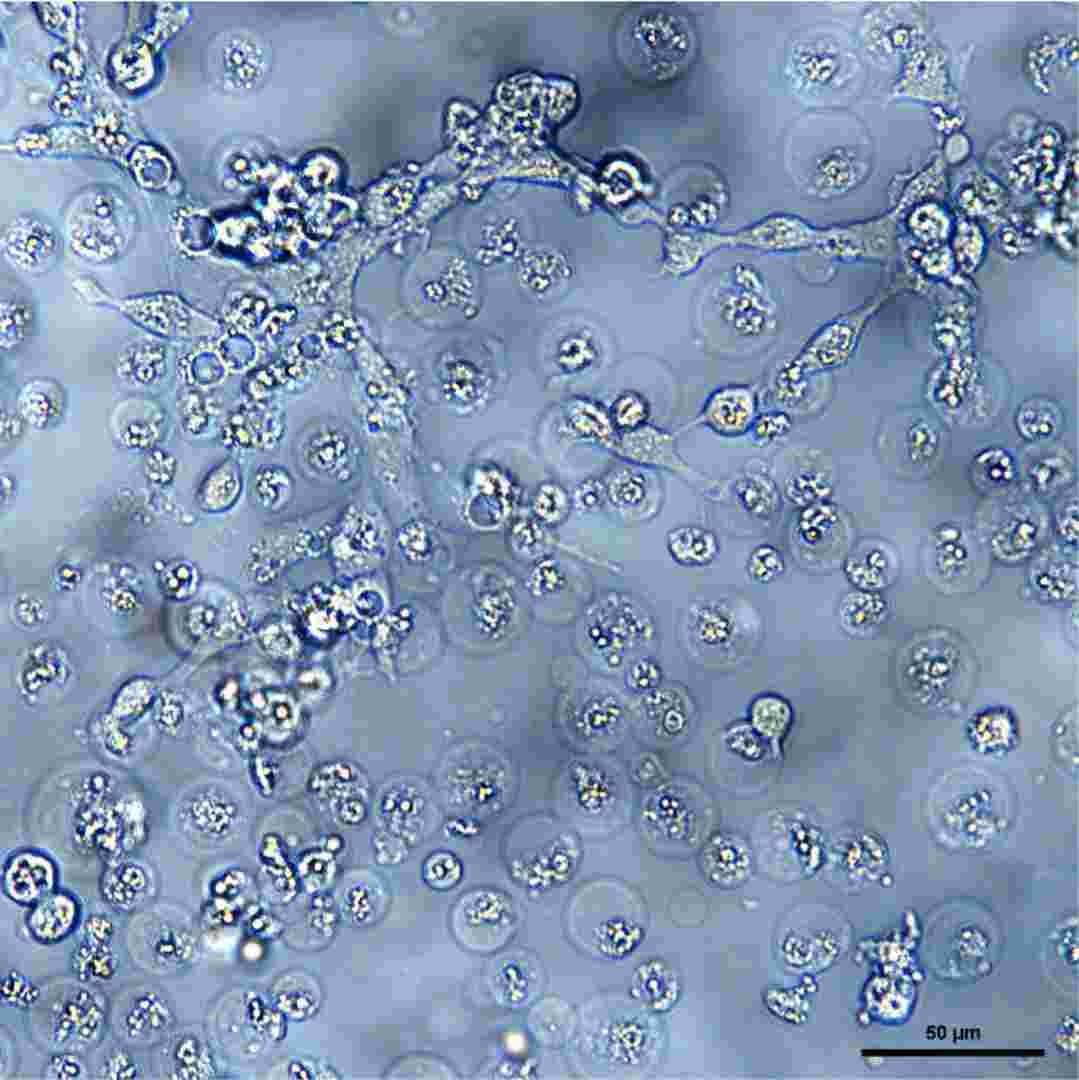 Fig. 2. Cytopathic effects in SSN-1 cells inoculated with the filtrate of naturally infected Murry cod fry tissue samples (Wang Y, Xu L, et al., 2021).
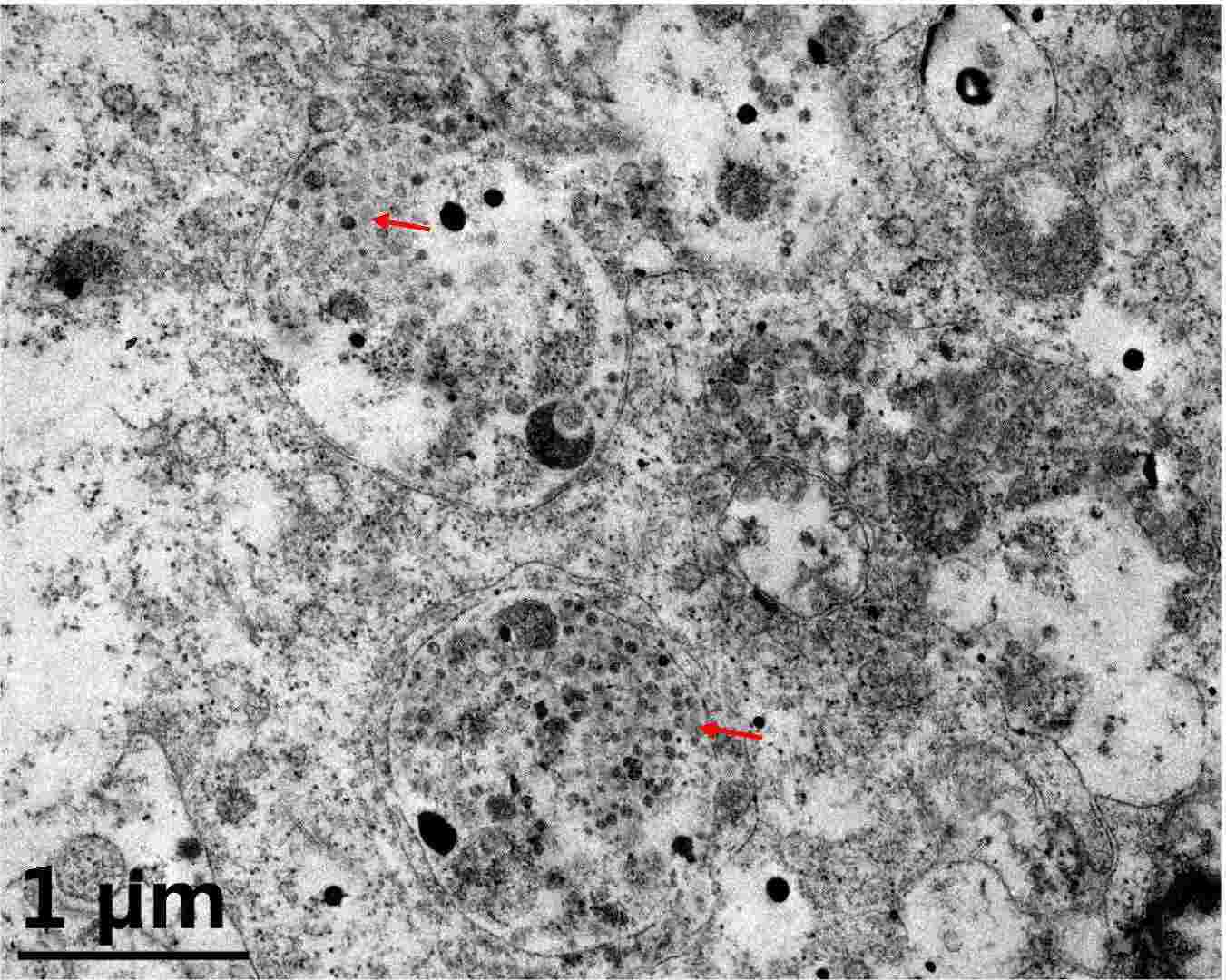 Fig. 3. Electron micrograph showing viral aggregates (arrows) within inclusion body of SSN-1 cells infected by the filtrate of naturally infected Murry cod fry tissue samples (Wang Y, Xu L, et al., 2021).
Ask a Question
Write your own review
- You May Also Need


Description: Derived from tissue posterior to the anus of normal adult minnows. Reported to be capable of growth over a wide temperature range from 4°C-36°C with maximum growth at 34°C. Support growth of the ...
Description: E11 is a clone of the cell line SSN-1 and is persistantly infected with a C-type retrovirus (SnRV). It is susceptible to piscine nodavirus strains belonging to different genotypes (SJNNV, RGNNV, ...

Description: From pooled male/female gonad tissue of yearling rainbow trout (Oncorhynchus mykiss). First cell line to be established from cold-blooded vertebrates. 8-fold difference in growth rate in temperature ...


- Adipose Tissue-Derived Stem Cells
- Human Neurons
- Mouse Probe
- Whole Chromosome Painting Probes
- Hepatic Cells
- Renal Cells
- In Vitro ADME Kits
- Tissue Microarray
- Tissue Blocks
- Tissue Sections
- FFPE Cell Pellet
- Probe
- Centromere Probes
- Telomere Probes
- Satellite Enumeration Probes
- Subtelomere Specific Probes
- Bacterial Probes
- ISH/FISH Probes
- Exosome Isolation Kit
- Human Adult Stem Cells
- Mouse Stem Cells
- iPSCs
- Mouse Embryonic Stem Cells
- iPSC Differentiation Kits
- Mesenchymal Stem Cells
- Immortalized Human Cells
- Immortalized Murine Cells
- Cell Immortalization Kit
- Adipose Cells
- Cardiac Cells
- Dermal Cells
- Epidermal Cells
- Peripheral Blood Mononuclear Cells
- Umbilical Cord Cells
- Monkey Primary Cells
- Mouse Primary Cells
- Breast Tumor Cells
- Colorectal Tumor Cells
- Esophageal Tumor Cells
- Lung Tumor Cells
- Leukemia/Lymphoma/Myeloma Cells
- Ovarian Tumor Cells
- Pancreatic Tumor Cells
- Mouse Tumor Cells