
GCT-27
Cat.No.: CSC-C6240X
Species: Homo sapiens (Human)
Source: Testis
Morphology: epitheloid cells growing adherently in monolayers in collagen-coated flasks
Culture Properties: monolayer
- Specification
- Background
- Scientific Data
- Q & A
- Customer Review
Immunology: cytokeratin +, cytokeratin-7 (+), cytokeratin-8 +, cytokeratin-17 -, cytokeratin-18 +, cytokeratin-19 +, desmin -, endothel -, EpCAM +, GFAP -, neurofilament -, vimentin +
Viruses: PCR: EBV -, HBV -, HCV -, HI
The human testicular germ cell tumor (TGCT) cell line GCT-27 (also known as GCT 27 or GCT27) was created from a patient who had a malignant testicular teratocarcinoma. It is a hypotriploid epithelial-type line that grows adherently in monolayers. Its sublines are described as nullipotent (GCT 27 C-1) and multipotent/pluripotent (GCT 27 X-1), the latter of which can differentiate into endodermal, ectodermal, and mesenchymal lineages when cultivated on feeder layers or when growth factors are removed.
GCT-27 is mostly utilized in the scientific literature as a model for germ cell tumor biology and human embryonal cancer. It has been widely used to investigate the processes regulating embryonal carcinoma cell differentiation vs self-renewal, the maintenance and loss of pluripotency, and the function of Oct4/SOX2/Nanog-related networks in germ cell neoplasia. Additionally, the line is a common tool in chemoresistance research, especially when examining acquired resistance pathways and cisplatin sensitivity in TGCTs, a characteristic of this illness. Furthermore, GCT-27 has been used in cytokine/growth factor signaling research, cancer testis antigen (CTA) expression profiling, and transcriptome investigations of solid tumors to distinguish between somatic and germ cell-specific oncogenic processes. Culturally, it is kept at 37°C and 5% CO2 in RPMI-1640 with serum supplementation.
Hyperactivation of the Fanconi Anemia Pathway Mediates Acquired Cisplatin Resistance in a Subset of Testicular Germ Cell Tumor Cells
Testicular germ cell tumors (TGCTs) are highly curable with cisplatin, yet ~20% of patients relapse or become refractory, with mechanisms of resistance remaining poorly understood. Using paired cisplatin-sensitive (cis-s) and -resistant (cis-r) TGCT cell lines, Caggiano et al. investigated the role of the Fanconi anemia (FA) DNA repair pathway in mediating acquired resistance.
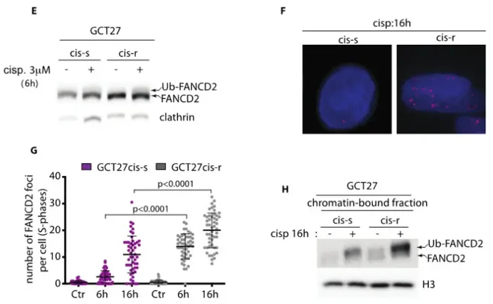
In GCT27cis-r cells, cisplatin exposure induced hyperactivation of the FA pathway compared to GCT27cis-s cells. Western blotting revealed increased basal FANCD2 expression and a greater fraction of monoubiquitinated FANCD2 (Ub-FANCD2) following drug treatment (Fig. 1E). Immunofluorescence confirmed a significantly higher number of nuclear FANCD2 foci in S-phase GCT27cis-r cells both during and after cisplatin exposure (Fig. 1F, G). This hyperactivation correlated with enhanced chromatin loading, as shown by increased Ub-FANCD2 in the chromatin-bound fraction (Fig. 1H).
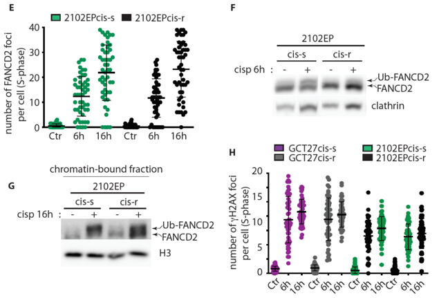
In contrast, the 2102EPcis-r model did not exhibit FA pathway hyperactivation. Despite increased basal FANCD2 expression (Fig. 2F), 2102EPcis-r cells showed no elevation in FANCD2 foci formation (Fig. 2E) or ubiquitination levels (Fig. 2G) compared to 2102EPcis-s cells. These findings demonstrate that hyperactivation of the FA repair pathway mediates cisplatin resistance in a subset of TGCT cells, highlighting mechanistic heterogeneity across resistant models.
Ask a Question
Write your own review
- You May Also Need
Description: Complex type germ cell tumor of human embryonic testis origin.
Description: Complex type germ cell tumor of human embryonic testis origin.
Description: A human embryonal carcinoma cell line derived from a human testicular germ cell tumour.
Description: NTERA-2 was cloned from cell line TERA-2 which was derived from a metastatic teratocarcinoma of a 22-year-old Caucasian male; cell line also known as NT-2
- Adipose Tissue-Derived Stem Cells
- Human Neurons
- Mouse Probe
- Whole Chromosome Painting Probes
- Hepatic Cells
- Renal Cells
- In Vitro ADME Kits
- Tissue Microarray
- Tissue Blocks
- Tissue Sections
- FFPE Cell Pellet
- Probe
- Centromere Probes
- Telomere Probes
- Satellite Enumeration Probes
- Subtelomere Specific Probes
- Bacterial Probes
- ISH/FISH Probes
- Exosome Isolation Kit
- Human Adult Stem Cells
- Mouse Stem Cells
- iPSCs
- Mouse Embryonic Stem Cells
- iPSC Differentiation Kits
- Mesenchymal Stem Cells
- Immortalized Human Cells
- Immortalized Murine Cells
- Cell Immortalization Kit
- Adipose Cells
- Cardiac Cells
- Dermal Cells
- Epidermal Cells
- Peripheral Blood Mononuclear Cells
- Umbilical Cord Cells
- Monkey Primary Cells
- Mouse Primary Cells
- Breast Tumor Cells
- Colorectal Tumor Cells
- Esophageal Tumor Cells
- Lung Tumor Cells
- Leukemia/Lymphoma/Myeloma Cells
- Ovarian Tumor Cells
- Pancreatic Tumor Cells
- Mouse Tumor Cells