CCD-1095Sk
Cat.No.: CSC-C9166W
Species: Homo sapiens (Human)
Source: Breast; Skin
Morphology: fibroblast
- Specification
- Background
- Scientific Data
- Q & A
- Customer Review
CCD-1095Sk is a human skin fibroblast cell line generated from normal skin tissue derived from a biopsy of the left breast of a 37-year-old woman with infiltrating ductal carcinoma and concomitant Paget's disease of the nipple. The cells were obtained from histologically normal skin, but from tissue close to a breast cancer lesion, so they provide a valuable model for the study of the behavior of stromal cells in the setting of the tumor microenvironment.
The cells display a normal fibroblast-like shape and develop as an adherent monolayer under standard culture conditions. Fibroblasts are cells of the connective tissue, which participate in the creation of the extracellular matrix, tissue remodeling and inter-cellular signaling. Thus, CCD-1095Sk cells provide an in vitro system to research fibroblast biology, cell-matrix interactions, wound repair mechanisms and stromal responses to external stimuli.
Unlike immortalized fibroblast models, CCD-1095Sk is a finite cell line that senesces after roughly 36 population doublings. This feature allows researchers to study cellular ageing, replicative senescence, and fibroblast functional changes over extended culture time. The cell line has also been reported to be suited for 3D culture applications, making it valuable for research of tissue architecture and stromal-epithelial interactions.
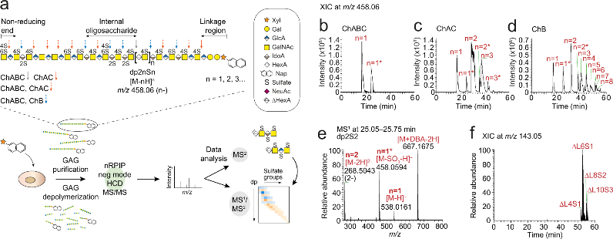
GAGDoMa: A High-Sensitivity Analytical Framework for Mapping the Glycosaminoglycome
Glycosaminoglycans (GAGs) are structurally diverse polysaccharides essential to numerous biological processes, yet comprehensive analysis of the "glycosaminoglycome" remains analytically challenging. To address this, Persson et al. developed GAGDoMa, a novel liquid chromatography-mass spectrometry (LC-MS) framework for high-resolution profiling of GAG oligosaccharides.
GAGDoMa employs bacterial lyases for controlled depolymerization: chondroitinase ABC fully degrades chondroitin sulfate/dermatan sulfate (CS/DS) into disaccharides and linkage-region structures, while chondroitinase AC and B cleave specifically at GlcA or IdoA residues, generating oligosaccharides of varying lengths. Heparinases selectively degrade heparan sulfate without affecting CS/DS. These eliminative reactions generate 4,5-unsaturated hexuronic acid (ΔHexA) residues, distinguishable by mass spectrometry from non-reducing end HexA residues (Δmass = 18.0106 Da) (Figure 1a). Xyloside-primed CS/DS was isolated from human breast fibroblasts (CCD-1095Sk) and breast carcinoma cells (HCC70), depolymerized, and analyzed using nanoflow reversed-phase ion-pairing (RPIP) chromatography with dibutylamine (DBA) as a volatile ion-pairing agent. Coupled to an LTQ Orbitrap Elite mass spectrometer in negative ionization mode, this setup achieved ~300-fold higher sensitivity and superior chromatographic resolution compared to previous microscale LC-MS/MS methods. Fragmentation was performed using stepped higher-energy collisional dissociation (HCD) at normalized collision energies (NCEs) of 20-80%, optimizing structural interpretation. Extracted ion chromatograms of the recurring precursor ion at m/z458.06-corresponding to internal oligosaccharides with one sulfate per disaccharide (dp2nSn)-revealed separation of species ranging from dp2S1 to dp16S8 (Figure 1b-d). Additionally sulfated oligosaccharides (dp2nSn+1) were detected and resolved chromatographically, despite in-source sulfate loss generating the same m/z458.06 signature (Figure 1e). Linkage-region structures (ΔL) containing the naphthyl aglycon were cleanly separated from internal oligosaccharides, enabling MS1-level identification (Figure 1f).
GAGDoMa enabled extensive compositional profiling of intact CS/DS, resolving species from L11S4 to L29S14 with mass accuracies <10 ppm. This framework provides unprecedented sensitivity and resolution for characterizing GAG structural diversity, offering a powerful tool for glycosaminoglycome mapping in biological and disease contexts.
Ask a Question
Write your own review
- You May Also Need
Description: Established in 2007 from the large retrosternal mass resected before treatment from a 57-year-old Caucasian man with rapidly fatal anaplastic thyroid cancer


Description: established from the uvea tissue of a male patient with uveal melanoma

Description: NTERA-2 was cloned from cell line TERA-2 which was derived from a metastatic teratocarcinoma of a 22-year-old Caucasian male; cell line also known as NT-2
Description: Established from the bone marrow of a 27-year-old woman with B-cell non-Hodgkin lymphoma (B-NHL) (diffuse large cell lymphoma, DLCL, stage 4B, at relapse) in 1987
- Adipose Tissue-Derived Stem Cells
- Human Neurons
- Mouse Probe
- Whole Chromosome Painting Probes
- Hepatic Cells
- Renal Cells
- In Vitro ADME Kits
- Tissue Microarray
- Tissue Blocks
- Tissue Sections
- FFPE Cell Pellet
- Probe
- Centromere Probes
- Telomere Probes
- Satellite Enumeration Probes
- Subtelomere Specific Probes
- Bacterial Probes
- ISH/FISH Probes
- Exosome Isolation Kit
- Human Adult Stem Cells
- Mouse Stem Cells
- iPSCs
- Mouse Embryonic Stem Cells
- iPSC Differentiation Kits
- Mesenchymal Stem Cells
- Immortalized Human Cells
- Immortalized Murine Cells
- Cell Immortalization Kit
- Adipose Cells
- Cardiac Cells
- Dermal Cells
- Epidermal Cells
- Peripheral Blood Mononuclear Cells
- Umbilical Cord Cells
- Monkey Primary Cells
- Mouse Primary Cells
- Breast Tumor Cells
- Colorectal Tumor Cells
- Esophageal Tumor Cells
- Lung Tumor Cells
- Leukemia/Lymphoma/Myeloma Cells
- Ovarian Tumor Cells
- Pancreatic Tumor Cells
- Mouse Tumor Cells