
SNG-M
Cat.No.: CSC-C6659J
Species: Homo sapiens (Human)
Source: Lymph Node Metastasis
Morphology: Polygonal Or Spindle
- Specification
- Background
- Scientific Data
- Q & A
- Customer Review
SNG-M is a well-characterized human endometrial adenocarcinoma cell line established from a metastatic lymph node specimen of a 52-year-old female patient. The line was established alongside its primary tumor counterpart, SNG-P, from the same patient. SNG-M exhibits a polygonal or spindle-shaped epithelial morphology, grows in a pavement arrangement with multilayering and no contact inhibition, and has demonstrated stable, uninterrupted growth for over 13 months. Karyotypically, the cells are aneuploid with a stable modal chromosome number in the diploid range. The line is negative for HPV types 16, 18, and 33 E6 oncogene sequences, confirming its HPV-independent origin.
A key advantage of SNG-M is its derivation from a metastatic site (lymph node), making it a valuable model for studying the molecular mechanisms of endometrial cancer invasion and dissemination. The cells are intermediately differentiated and have been extensively utilized in investigations of steroid hormone responses. SNG-M exhibits growth suppression and differentiation induction upon progesterone treatment, both in vitro and in vivo. Furthermore, its migration and invasion into Matrigel are inhibited by medroxyprogesterone acetate (MPA) and danazol, but promoted by EGF and TGF-α in a concentration-dependent manner. Notably, SNG-M demonstrates temperature sensitivity, with a permissive growth temperature approximately 3°C lower than other endometrial lines such as HAIN, and ceases growth at temperatures above 39.5°C.
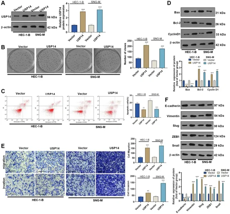
Overexpressing USP14 Reduced Apoptosis and Promoted EC Cell Migration and Invasion
Ubiquitin-specific protease 14 (USP14), a member of the USP family, which catalyzes ubiquitin cleavage from a range of protein substrates, has been found dysregulated in several cancers. We transfected USP14 overexpression plasmids into HEC-1-B and SNG-M cells to clarify the specific mechanism of USP14 in endometrial carcinoma (EC). First, WB showed that USP14 was highly expressed in EC cells, indicating successful transfection (Figure 1A). The assessment of EC cell proliferation and apoptosis was performed using the colony formation assay and FCM, respectively. The results revealed that upregulating USP14 facilitated the colony-forming ability of HEC-1-B and SNG-M and reduced apoptosis (vs. the vector group, Figure 1B, 1C). Furthermore, apoptosis-related proteins (Bcl-2, Bax, and Cyclin D1) were compared by WB. As a result, overexpressing USP14 dampened the Bax level and enhanced the Bcl-2 and Cyclin D1 profiles (Figure 1D). Subsequently, the Transwell experiment assessed the impact of USP14 on EC cell migratory and invasive capabilities. Interestingly, cell migration and invasion were improved after USP14 overexpression (vs. the vector group) (Figure 1E). In addition, the profiles of EMT-related proteins (E-cadherin and Vimentin) and EMT-related transcription factors (Snail1, ZEB1, and Slug) were compared by WB. USP14 overexpression elevated the profiles of Vimentin, Snail1, ZEB1, and Slug while repressing the E-cadherin level (compared with the vector group, Figure 1F). These findings illustrated that overexpressing USP14 facilitated EC cell migration, invasion, and EMT.
Ask a Question
Write your own review
- You May Also Need
Description: Uterine adenosquamous carcinoma. Said CEA and CA125 producing. Cell growth is slow.
Description: Established from tumor tissue from a 77-year-old woman with recurrence of endometrial carcinoma (adenomatous, partly papillary, grade G3) in 1990; described as forming heterotransplantable tumors in ...
Description: Glassy cell carcinoma. TA-4, CA125, neuron-specific enolase producing.
Description: The cells possess alpha keratin, well defined junctional complexes, tonofilaments and surface microvilli.
- Adipose Tissue-Derived Stem Cells
- Human Neurons
- Mouse Probe
- Whole Chromosome Painting Probes
- Hepatic Cells
- Renal Cells
- In Vitro ADME Kits
- Tissue Microarray
- Tissue Blocks
- Tissue Sections
- FFPE Cell Pellet
- Probe
- Centromere Probes
- Telomere Probes
- Satellite Enumeration Probes
- Subtelomere Specific Probes
- Bacterial Probes
- ISH/FISH Probes
- Exosome Isolation Kit
- Human Adult Stem Cells
- Mouse Stem Cells
- iPSCs
- Mouse Embryonic Stem Cells
- iPSC Differentiation Kits
- Mesenchymal Stem Cells
- Immortalized Human Cells
- Immortalized Murine Cells
- Cell Immortalization Kit
- Adipose Cells
- Cardiac Cells
- Dermal Cells
- Epidermal Cells
- Peripheral Blood Mononuclear Cells
- Umbilical Cord Cells
- Monkey Primary Cells
- Mouse Primary Cells
- Breast Tumor Cells
- Colorectal Tumor Cells
- Esophageal Tumor Cells
- Lung Tumor Cells
- Leukemia/Lymphoma/Myeloma Cells
- Ovarian Tumor Cells
- Pancreatic Tumor Cells
- Mouse Tumor Cells