
MFE-280
Cat.No.: CSC-C0454
Species: Homo sapiens (Human)
Source: Uterus; Endometrium
Morphology: epitheloid cells growing adherently in monolayers
Culture Properties: monolayer
- Specification
- Background
- Scientific Data
- Q & A
- Customer Review
Immunology: cytokeratin +, cytokeratin-7 -, cytokeratin-8 +, cytokeratin-17 -, cytokeratin-18 +, cytokeratin-19 +, desmin -, endothel -, EpCAM +, GFAP -, neurofilament -,
MFE-280 was developed from a tumor specimen from a 77-year-old woman diagnosed with recurrent endometrial adenocarcinoma, adenomatous, with focal papillary features, G3. Endometrial cancer is the most common cancer of the female reproductive system in postmenopausal women. MFE-280 has a hyperdiploid karyotype, 48–52 chromosomes with many chromosomal abnormalities (-X, +1 (x2), -2, +9, -10, -12, -13, -16, -17 (x2), -18, -19, -20, -22, and +18mar). These chromosomal anomalies include common genetic changes in endometrial cancer such as gain of chromosome arm 1q. Additionally, MFE-280 cells express the progesterone receptor, suggesting potential sensitivity to hormone-based therapies. MFE-280 cells have epithelial-like morphology. They grow in an adherent manner, the cells are round or irregularly polygonal with tight junctions between cells, all features typical for epithelial cells.
Research has utilized the MFE-280 cell line to model endometrial cancer through various experimental studies including drug testing. For example, shikonin has been shown to inhibit MFE-280 cell proliferation, indicating its potential as an anti-cancer compound. Moreover, overexpression of miR-124-3p has been shown to affect the biological behavior of MFE-280 cells and therefore could have therapeutic potential in endometrial cancer.
MSM Decreases Viability of EC Cells in a Dose-Dependent Manner
Methylsulfonylmethane (MSM) is an over-the-counter dietary supplement with anti-inflammatory, antioxidative, and osteoarthritis, gastric injury, obesity-associated disorders, and other conditions ameliorating properties. Multiple research studies have demonstrated that MSM holds therapeutic potential for treating various cancer types such as endometrial cancer (EC). In this study, Kowalska et al. used EC cell lines (ISHIKAWA, MFE-296, and MFE-280) to determine the effects of MSM, either alone or in combination with the chemotherapy drug doxorubicin (DOX).
Firstly, they evaluated the effect of MSM on EC cells and observed that MSM decreases the viability of EC cells in a dose-dependent manner (Fig. 1a). ISHIKAWA cell line, which represents the well differentiated EC, presented the highest sensitivity to MSM as compared to moderately differentiated MFE-296 and poorly differentiated MFE-280. A significant decrease in the viability of ISHIKAWA and MFE-296 cell lines was observed for doses of MSM below 600 mM. MFE-280 cells demonstrated a significant response at 300 mM and higher doses. For the rest of experiments, the concentrations of 300 mM and 400 mM of MSM were chosen. Both doses of MSM are for most of the cell lines above the IC50: for ISHIKAWA IC50 = 538.6 mM, MFE-296 IC50 = 551.1 mM MSM, and for MFE-280 IC50 = 380 mM MSM, based on AlamarBlue assay results.
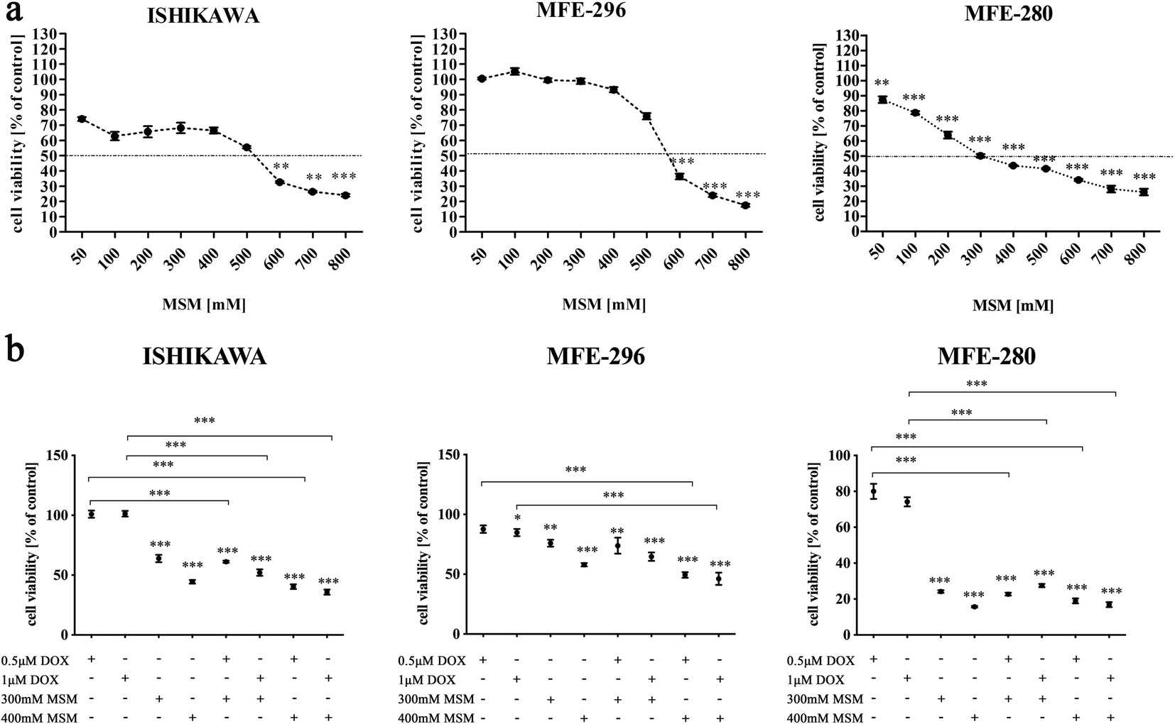 Fig. 1. Methylsulfonylmethane sensitizes endometrial cancer cells to doxorubicin (Kowalska K, Habrowska-Górczyńska D E, et al., 2021).
Fuling Overrides the Estradiol-Mediated Potentiation of Endometrial Cancer Cell Lines Invasiveness
The Traditional Chinese medicine, Guizhi Fuling (here called Fuling), has been confirmed in meta-analysis studies to reduce recurrence of endometriosis and improve pregnancy outcomes; however, the possible use of Fuling as a fertility-preserving treatment in endometrial cancer has not previously been tested. Khan et al. explored this by testing Fuling on two endometrial cancer cell lines (Ishikawa and MFE-280).
The extract of Fuling inhibited the invasiveness of both cell lines in a dose-dependent manner. The invasiveness of cells was assessed using transwell assays after 24 hours (Fig. 2). Box plots show that Fuling decreased invasiveness by 50% at around 58 µg/ml for Ishikawa and 52 µg/ml for MFE-280 (Fig. 2A and B). Representative invasion images are presented in Figure 2C. In order to evaluate its effects on 2D surface migration, a wound was scratched in confluent cell monolayers. After 24 hours of incubation with 80 µg/ml of Fuling or vehicle (DMSO), wound closure was calculated with NIH-Image software. Fuling significantly reduced migration in both cell lines when compared to control (Fig. 3). The images of wound closure are shown in Figure 3A, B, and percent closure was calculated by normalizing to wound area before treatment (Fig. 3C).
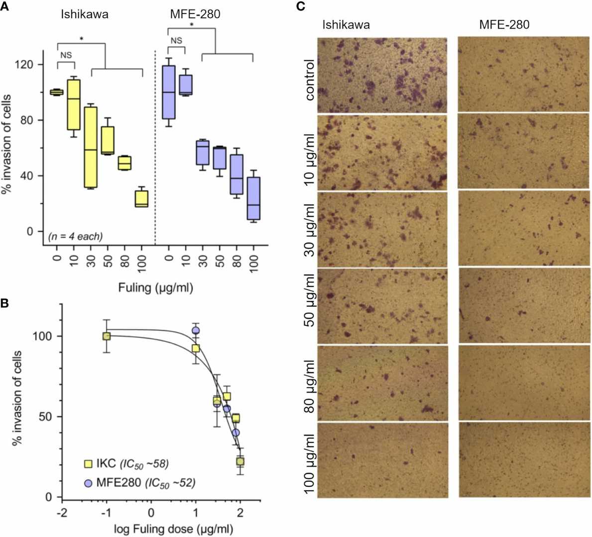 Fig. 2. Dose-dependent inhibition of invasion of Ishikawa and MFE-280 cells by Fuling (Khan S, Varricchio A, et al., 2022).
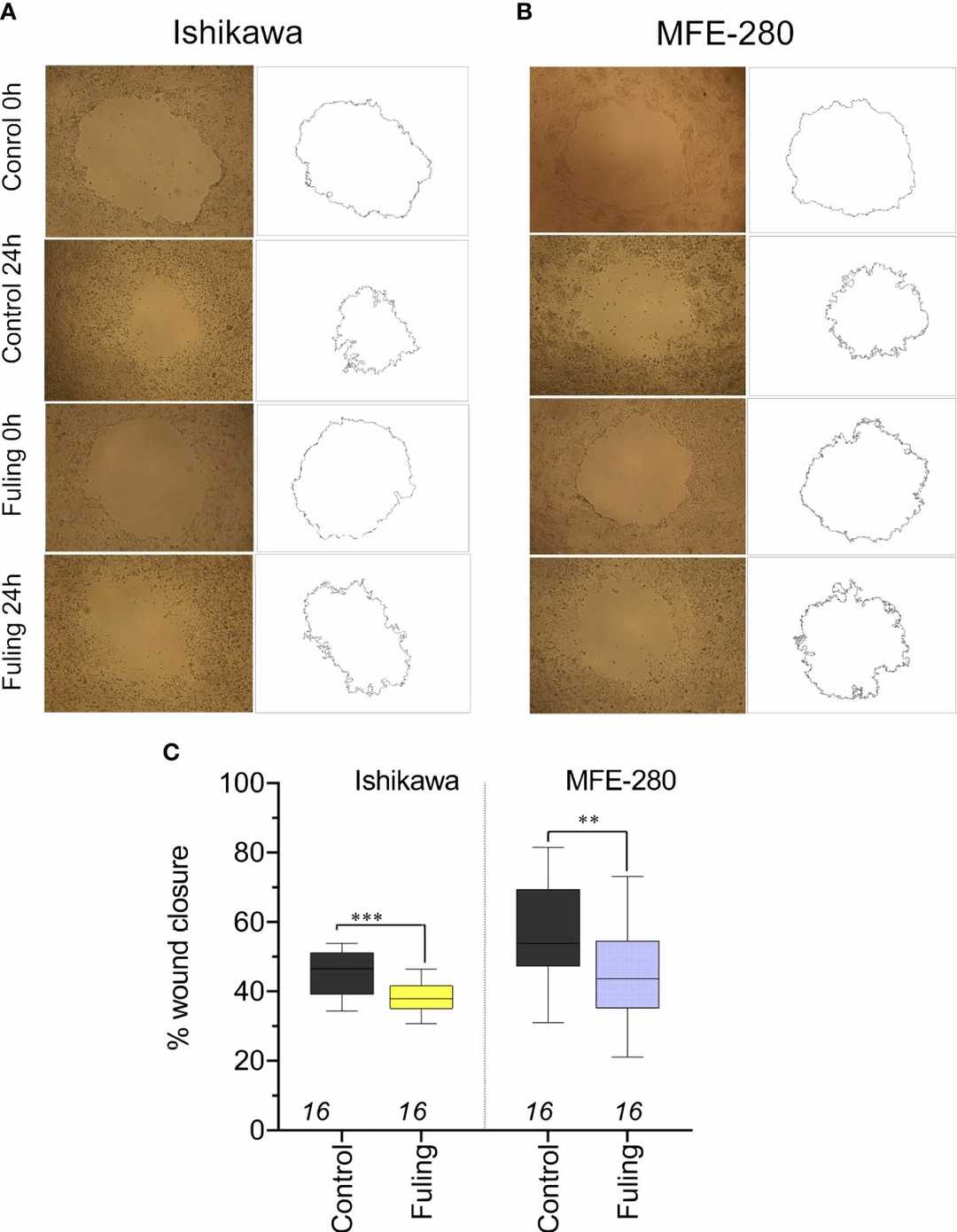 Fig. 3. Decreased motility measured by reduced wound closure rates in plated Ishikawa and MFE-280 in cultures treated with 80 µg/ml Fuling, as compared with vehicle control (Khan S, Varricchio A, et al., 2022).
Ask a Question
Write your own review
- You May Also Need

Description: Uterine adenosquamous carcinoma. Said CEA and CA125 producing. Cell growth is slow.

Description: Glassy cell carcinoma. TA-4, CA125, neuron-specific enolase producing.
Description: The cells possess alpha keratin, well defined junctional complexes, tonofilaments and surface microvilli.


Description: Established from the ascitic fluid of a 69-year-old Caucasian woman with endometrial carcinoma in relapse in 1985
- Adipose Tissue-Derived Stem Cells
- Human Neurons
- Mouse Probe
- Whole Chromosome Painting Probes
- Hepatic Cells
- Renal Cells
- In Vitro ADME Kits
- Tissue Microarray
- Tissue Blocks
- Tissue Sections
- FFPE Cell Pellet
- Probe
- Centromere Probes
- Telomere Probes
- Satellite Enumeration Probes
- Subtelomere Specific Probes
- Bacterial Probes
- ISH/FISH Probes
- Exosome Isolation Kit
- Human Adult Stem Cells
- Mouse Stem Cells
- iPSCs
- Mouse Embryonic Stem Cells
- iPSC Differentiation Kits
- Mesenchymal Stem Cells
- Immortalized Human Cells
- Immortalized Murine Cells
- Cell Immortalization Kit
- Adipose Cells
- Cardiac Cells
- Dermal Cells
- Epidermal Cells
- Peripheral Blood Mononuclear Cells
- Umbilical Cord Cells
- Monkey Primary Cells
- Mouse Primary Cells
- Breast Tumor Cells
- Colorectal Tumor Cells
- Esophageal Tumor Cells
- Lung Tumor Cells
- Leukemia/Lymphoma/Myeloma Cells
- Ovarian Tumor Cells
- Pancreatic Tumor Cells
- Mouse Tumor Cells