
PACADD-159
Cat.No.: CSC-C6237X
Species: Homo sapiens (Human)
Source: Pancreas
Morphology: epitheloid cells growing adherently in monolayers
Culture Properties: monolayer
- Specification
- Background
- Scientific Data
- Q & A
- Customer Review
Immunology: cytokeratin +, desmin -, endothel -, GFAP -, neurofilament -, vimentin -
Viruses: PCR: EBV -, HBV -, HCV -, HIV -, HTLV-I/-II -
PACADD-159 (aka PaCaDD-159) is a pancreatic cancer cell line from a pancreatic ductal adenocarcinoma (PDAC) patient tumor. Specifically, the original tumor was a primary PDAC tumor from a male who was 78 years old at the time of diagnosis/tumor procurement. It was derived by the Dresden outgrowth protocol and created as part of a collection of primary PDAC tumor cell lines with the intent of modeling tumor heterogeneity and clinical pathology more accurately. It has epithelial like morphology and forms adherent monolayer cultures. PACADD-159 has slower growth compared to other pancreatic cancer cells lines and poor ability to form 3D tumor spheres, indicating possible higher microenvironment dependency.
As anticipated for a pancreatic cancer cell line, it carries oncogenic KRAS mutations. Transcriptomic analysis has shown upregulation of NF-κB, PI3K-Akt signaling, and genes involved in tumor progression, EMT, and drug resistance. The cell line has recently been used for differential transcriptomic and proteomic profiling comparing tumor heterogeneity, drug response differences, and secretome analysis between pancreatic cancer cell lines.
Growth Characteristics of Primary and Expandable Pancreatic Cancer Cell Lines
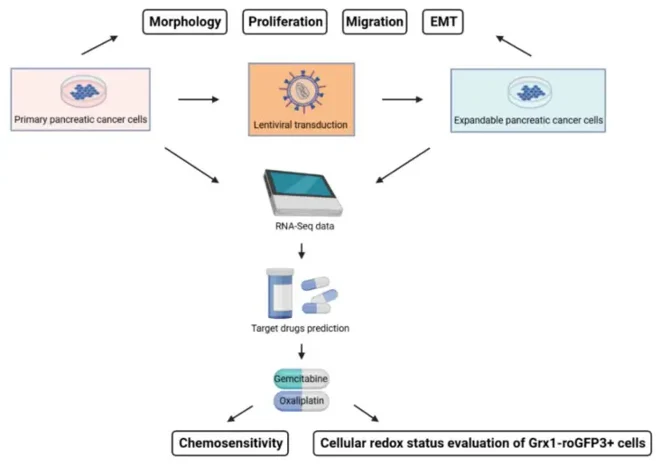
Guo's team addresses the challenge that primary pancreatic cancer cells become highly vulnerable and difficult to maintain after losing their tumor microenvironment. They investigated whether lentiviral transduction could generate expandable primary pancreatic cancer cell lines retaining original tumor-specific characteristics, including chemosensitivity, for patient-specific chemotherapy screening.
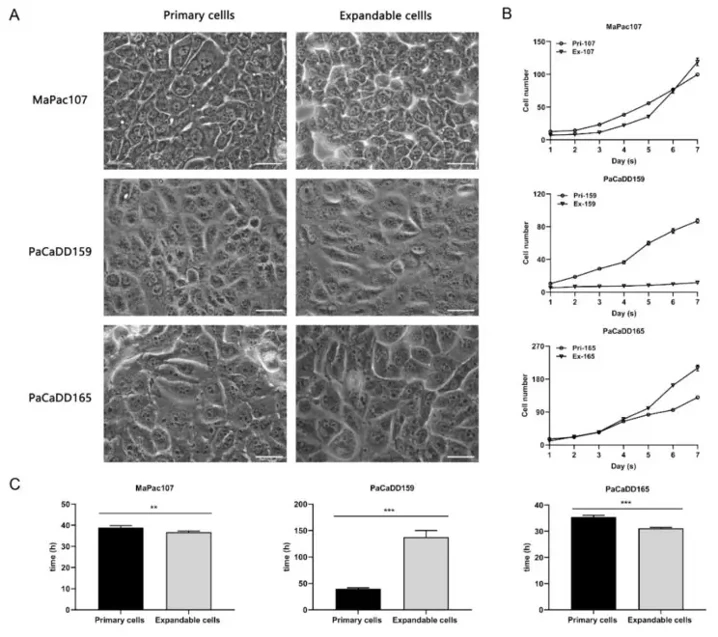
Figure 1 shows the study flowchart. Cell morphology was assessed in 2D culture (Figure 2A). Primary MaPac107 (Pri-MaPac107) exhibited epithelial monolayer characteristics with a fusiform-organized pattern, growing in clusters before confluency with irregularly shaped nuclei; this pattern disappeared at confluence. Expandable MaPac107 (Ex-MaPac107) shared identical morphology. Primary PaCaDD159 (Pri-PaCaDD159) proliferated as epithelial monolayers with lumpy, cluster-like organization, featuring ovoid cells with small round nuclei; Ex-PaCaDD159 showed matching morphology. Primary PaCaDD165 (Pri-PaCaDD165) grew as small polygonal cells with a cobblestone pattern, prominent nuclei, and rapid proliferation; Ex-PaCaDD165 showed no morphological differences.
Growth curves and doubling times are shown in Figure 2B, C. Ex-MaPac107 and Ex-PaCaDD165 exhibited higher proliferation rates than their primary counterparts, while Ex-PaCaDD159 proliferated significantly slower than Pri-PaCaDD159. Despite this unexpected reduction, the remaining two expandable lines demonstrated enhanced proliferation in 2D culture.
Ask a Question
Write your own review

Description: Human moderately differentiated adenocarcinoma cell line established from liver metastasis.

Description: Established in 1985 from the primary ductal pancreatic adenocarcinoma (grade II) from a 44-year-old woman; reported as inducing metastasis in nude mice and as secreting proteinases (e.g. urokinase, ...

Description: established from the pancreas of a 81-year-old male patient with pancreatic ductal adenocarcinoma

Description: The MIA PaCa-2 cell line was established from tumor tissue of the pancreas obtained from a 65-year-old Caucasian male in 1975 by A. Yunis, et al. These cells are described to express human colony ...

Description: Established in 2010 from the pancreatic tumor of a 46-year old man with poorly differentiated ductal adenocarcinoma (high grade G3, pT3pN1)

Description: Human tumor cell line secreting parathyroid hormone-related peptide (PTHrP) established from pancreatic tumor.
- Adipose Tissue-Derived Stem Cells
- Human Neurons
- Mouse Probe
- Whole Chromosome Painting Probes
- Hepatic Cells
- Renal Cells
- In Vitro ADME Kits
- Tissue Microarray
- Tissue Blocks
- Tissue Sections
- FFPE Cell Pellet
- Probe
- Centromere Probes
- Telomere Probes
- Satellite Enumeration Probes
- Subtelomere Specific Probes
- Bacterial Probes
- ISH/FISH Probes
- Exosome Isolation Kit
- Human Adult Stem Cells
- Mouse Stem Cells
- iPSCs
- Mouse Embryonic Stem Cells
- iPSC Differentiation Kits
- Mesenchymal Stem Cells
- Immortalized Human Cells
- Immortalized Murine Cells
- Cell Immortalization Kit
- Adipose Cells
- Cardiac Cells
- Dermal Cells
- Epidermal Cells
- Peripheral Blood Mononuclear Cells
- Umbilical Cord Cells
- Monkey Primary Cells
- Mouse Primary Cells
- Breast Tumor Cells
- Colorectal Tumor Cells
- Esophageal Tumor Cells
- Lung Tumor Cells
- Leukemia/Lymphoma/Myeloma Cells
- Ovarian Tumor Cells
- Pancreatic Tumor Cells
- Mouse Tumor Cells