
N1E-115-1
Cat.No.: CSC-C9409J
Species: Mus musculus (Mouse)
Source: Peripheral Nervous System
- Specification
- Background
- Scientific Data
- Q & A
- Customer Review
N1E-115-1 is a subclone of the mouse neuroblastoma cell line N1E-115 first generated by Amano et al. In the typical culture conditions, the N1E-115-1 cells grow with a rather undifferentiated appearance. Under serum-reduced or differentiation-inducing circumstances, the cells are able to extend neurite-like processes and acquire neuron-like properties, thereby providing an ideal in vitro model for research of neuronal differentiation and neurite outgrowth.
N1E-115-1 cells are often employed to study signaling pathways involved in neuronal development, cytoskeletal architecture, intracellular trafficking, and neurotrophin responses. The cells respond to growth factors such as nerve growth factor (NGF), brain-derived neurotrophic factor (BDNF) and neurotrophin-3 (NT-3) allowing researchers to investigate molecular mechanisms controlling neurite production and elongation. Recent investigations using gene-editing N1E-115 cells have showed that loss of the SNARE proteins Vti1a and Vti1b affects neuronal differentiation, neurite outgrowth and Akt and ERK signaling pathways after neurotrophin stimulation. These results support the use of N1E-115-derived models to investigate mechanisms in neuronal signaling and membrane trafficking.
N1E-115 cells have been used also in oxidative stress and neurodegenerative research. Hydrogen peroxide at low concentrations leads to tau hyperphosphorylation through microtubule affinity-regulating kinases (MARKs) activation. This system provides an experimental model for studying cellular mechanisms concerning tau disease.
Live Labeling of the Axon Initial Segment via Unnatural Amino Acid Incorporation
The axon initial segment (AIS) is a specialized neuronal domain critical for action potential initiation and polarity maintenance. To enable live imaging of AIS components, Stajković et al. developed a method using unnatural amino acid (UAA) incorporation and click chemistry. This approach leverages the small size of UAAs and their flexible insertion sites to label large, structurally complex proteins without disrupting function.
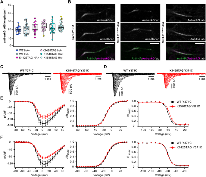
They successfully labeled two major AIS proteins in primary neurons: the 186 kDa neurofascin-186 (NF186) and the 260 kDa voltage-gated sodium channel NaV1.6 (encoded by Scn8a), using conventional and super-resolution microscopy. To validate the technique, they assessed whether UAA tagging altered AIS structure or channel biophysics. Overexpression of clickable NaV1.6 variants did not significantly affect AIS length compared to untransfected neighboring neurons (Fig. 1A, B).
Electrophysiological analysis using whole-cell patch clamp in N1E-115-1 cells revealed that incorporation of the UAA (TCOA-Lys) had minimal impact on channel function. The NaV1.6K1546TAG variant caused a small but significant depolarizing shift (+2.8 mV) in the fast inactivation curve, slightly slowed inactivation kinetics, and accelerated recovery (Fig. 1C, E). For the NaV1.6K1425TAG variant, peak sodium current density was reduced compared to wild-type, while other gating parameters remained unchanged (Fig. 4D, F*). Transient transfection efficiency was improved by using a PiggyBac transposase system to stably integrate β-subunits and a P2A-linked eGFP reporter, enabling reliable recording of larger currents.
These results demonstrate that UAA-mediated click labeling provides a robust, minimally invasive strategy for live visualization and functional analysis of AIS proteins, facilitating studies of neuronal excitability and polarity.
Ask a Question
Write your own review
Description: Described as secreting a mouse monoclonal antibody (IgG2a) detecting all fibers in skeletal muscle and myosin heavy chains on Western blots and detecting mammalian, chicken, zebrafish, axolotl, ...
Description: Animals were immunized with the B6.1 mouse cytotoxic T cell line.
Description: neuroglial and neuronal character coexpressing ependymoma cell line.
Description: Established by irradiation of the adherent cells in long-term bone marrow cultures derived from C3H/HeNSlc strain mice
- Adipose Tissue-Derived Stem Cells
- Human Neurons
- Mouse Probe
- Whole Chromosome Painting Probes
- Hepatic Cells
- Renal Cells
- In Vitro ADME Kits
- Tissue Microarray
- Tissue Blocks
- Tissue Sections
- FFPE Cell Pellet
- Probe
- Centromere Probes
- Telomere Probes
- Satellite Enumeration Probes
- Subtelomere Specific Probes
- Bacterial Probes
- ISH/FISH Probes
- Exosome Isolation Kit
- Human Adult Stem Cells
- Mouse Stem Cells
- iPSCs
- Mouse Embryonic Stem Cells
- iPSC Differentiation Kits
- Mesenchymal Stem Cells
- Immortalized Human Cells
- Immortalized Murine Cells
- Cell Immortalization Kit
- Adipose Cells
- Cardiac Cells
- Dermal Cells
- Epidermal Cells
- Peripheral Blood Mononuclear Cells
- Umbilical Cord Cells
- Monkey Primary Cells
- Mouse Primary Cells
- Breast Tumor Cells
- Colorectal Tumor Cells
- Esophageal Tumor Cells
- Lung Tumor Cells
- Leukemia/Lymphoma/Myeloma Cells
- Ovarian Tumor Cells
- Pancreatic Tumor Cells
- Mouse Tumor Cells