MLg
Cat.No.: CSC-C9496L
Species: Mus musculus (Mouse)
Source: Lung
Morphology: Fibroblast-like
Culture Properties: adherent
- Specification
- Background
- Scientific Data
- Q & A
- Customer Review
MLg is an immortalized mouse lung fibroblast cell line that has been commonly utilized as a non-transformed stromal/mesenchymal cell model in cell biology, pulmonary research, and extracellular matrix studies. Isolated from the lung of a young C3H/He mouse, MLg cells display typical fibroblast morphology characterized by an elongated spindle-shape with a propensity for adherent growth on tissue culture plastic. MLg cells propagate as a mostly homogenous monolayer of cells with an intermediate doubling time when maintained under conventional cell culture conditions (e.g. DMEM supplemented with 10% fetal bovine serum) and express canonical fibroblast markers such as vimentin and fibroblast surface protein (FSP) without evidence of epithelial or endothelial cell marker expression. For these reasons MLg cells have been used to study cell-matrix interactions, mechano-transduction, fibroblast proliferation and differentiation, and stromal-epithelial communication using co-culture models.
MLg cells have been used in numerous pulmonary fibrosis, wound healing, and respiratory toxicology studies, where researchers have investigated fibroblast activation/myofibroblast trans-differentiation, collagen/elastin production, and responsiveness to profibrotic cytokines such as TGF-β1. MLg cells have also been used to study extracellular matrix remodeling, cellular senescence, oxidative stress, and numerous mesenchymal signal transduction pathways (e.g., MAPK, PI3K/AKT).
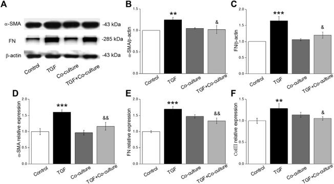
BMSCs Rescues TGF-β1-Induced Myofibroblast Differentiation In Vitro
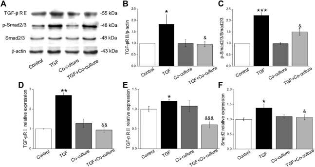
Pulmonary fibrosis (PF) is a chronic progressive interstitial lung disease with poor prognosis, driven by abnormal TGF-β1 activation and fibroblast differentiation. Liu's team investigated bone marrow-derived MSC (BMSC) effects on TGF-β1-induced fibrosis using co-cultures with mouse lung fibroblasts (MLg).
They found that stimulation with TGF-β1 (10 ng/mL, 24h) could establish a PF model, which upregulated fibrosis markers α-SMA and FN. Compared with PF cells, BMSC co-culture abrogated the increased expression levels of α-SMA and FN (Fig. 1A-C). qRT-PCR analysis showed the same results (Fig. 1D-F). These results suggested that BMSCs attenuate TGF-β1-induced myofibroblast differentiation. They also found that TGF-β/Smad pathway related proteins expression was upregulated by TGF-β1 treatment (10 ng/mL, 24h) and co-culture with BMSC reduced their expression (Fig. 2A-C). qRT-PCR assays further confirmed this result at the mRNA level (Fig. 2D-F). Taken together, TGF-β1/Smad signaling pathway is involved in BMSC treatment against PF.
Ask a Question
Write your own review
- You May Also Need

Description: Described as secreting a mouse monoclonal antibody (IgG2a) detecting all fibers in skeletal muscle and myosin heavy chains on Western blots and detecting mammalian, chicken, zebrafish, axolotl, ...

Description: Animals were immunized with the B6.1 mouse cytotoxic T cell line.


Description: neuroglial and neuronal character coexpressing ependymoma cell line.

Description: Established by irradiation of the adherent cells in long-term bone marrow cultures derived from C3H/HeNSlc strain mice
- Adipose Tissue-Derived Stem Cells
- Human Neurons
- Mouse Probe
- Whole Chromosome Painting Probes
- Hepatic Cells
- Renal Cells
- In Vitro ADME Kits
- Tissue Microarray
- Tissue Blocks
- Tissue Sections
- FFPE Cell Pellet
- Probe
- Centromere Probes
- Telomere Probes
- Satellite Enumeration Probes
- Subtelomere Specific Probes
- Bacterial Probes
- ISH/FISH Probes
- Exosome Isolation Kit
- Human Adult Stem Cells
- Mouse Stem Cells
- iPSCs
- Mouse Embryonic Stem Cells
- iPSC Differentiation Kits
- Mesenchymal Stem Cells
- Immortalized Human Cells
- Immortalized Murine Cells
- Cell Immortalization Kit
- Adipose Cells
- Cardiac Cells
- Dermal Cells
- Epidermal Cells
- Peripheral Blood Mononuclear Cells
- Umbilical Cord Cells
- Monkey Primary Cells
- Mouse Primary Cells
- Breast Tumor Cells
- Colorectal Tumor Cells
- Esophageal Tumor Cells
- Lung Tumor Cells
- Leukemia/Lymphoma/Myeloma Cells
- Ovarian Tumor Cells
- Pancreatic Tumor Cells
- Mouse Tumor Cells