KYSE-270
Cat.No.: CSC-C0428
Species: Homo sapiens (Human)
Source: Esophagus
Morphology: epitheloid cells growing in monolayers
Culture Properties: monolayer
- Specification
- Background
- Scientific Data
- Q & A
- Customer Review
Immunology: cytokeratin +, cytokeratin-7 - , cytokeratin-8 +, cytokeratin-17 (+), cytokeratin-18 +,
The KYSE-270 cell line is a well-characterized human cellular model derived from a primary esophageal squamous cell carcinoma (ESCC). ESCC is a major global health burden and the predominant histological subtype of esophageal cancer. KYSE-270, therefore, represents a highly relevant in vitro and in vivo system for studying the pathogenesis, progression, and therapeutic vulnerabilities of this aggressive malignancy. Its derivation from a primary tumor ensures it captures the genetic and phenotypic landscape of the disease at a clinically significant stage.
KYSE-270 cells exhibit typical epithelial morphology. A defining molecular feature is the presence of a nonsense mutation in the TP53 tumor suppressor gene (R213*), which is one of the most frequently mutated genes in ESCC. This loss of functional p53 contributes to genomic instability and impaired apoptosis, key hallmarks of cancer. The cell line is tumorigenic in immunocompromised mice, capable of forming solid tumors, which is essential for translational preclinical studies. It is characterized by a moderately differentiated phenotype and has been extensively profiled for its genomic, transcriptomic, and drug-response characteristics, making it a well-annotated tool in the ESCC research community.
CRISPR/Cas9 Screening Highlights PFKFB3 Gene as a Major Contributor to 5-Fluorouracil Resistance in Esophageal Cancer
Esophageal cancer (EC) is the eighth most common cancer and the sixth leading cause of death worldwide. Esophageal squamous cell carcinoma (ESCC) comprises the majority of esophageal cancers globally, and 5-Fluorouraci (5-FU) is one of the commonly used chemotherapeutics for this type of cancer. Chemoresistance is a major obstacle to the successful treatment of this malignancy.
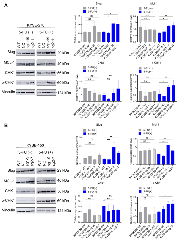
This study used CRISPR/Cas9 screening method to determine target genes related to 5-FU drug resistance in esophageal cancer. The results showed that loss of PFKFB3 can increase 5-FU resistance in different human esophageal squamous cell carcinoma cell lines. Specifically, in KYSE-70 cells, loss of PFKFB3 induce epithelial-mesenchymal transition (EMT) and prolong the S phase of the cell cycle, enabling cancer cells to evade the effects of 5-FU and develop resistance. In KYSE-270 and KYSE-150 cell lines, loss of PFKFB3 upregulate the expression of Slug and Mcl-1, indirectly regulate Chk1 and promote its autophosphorylation, which in turn inhibits apoptosis, thus counteracting the effects of 5-FU.
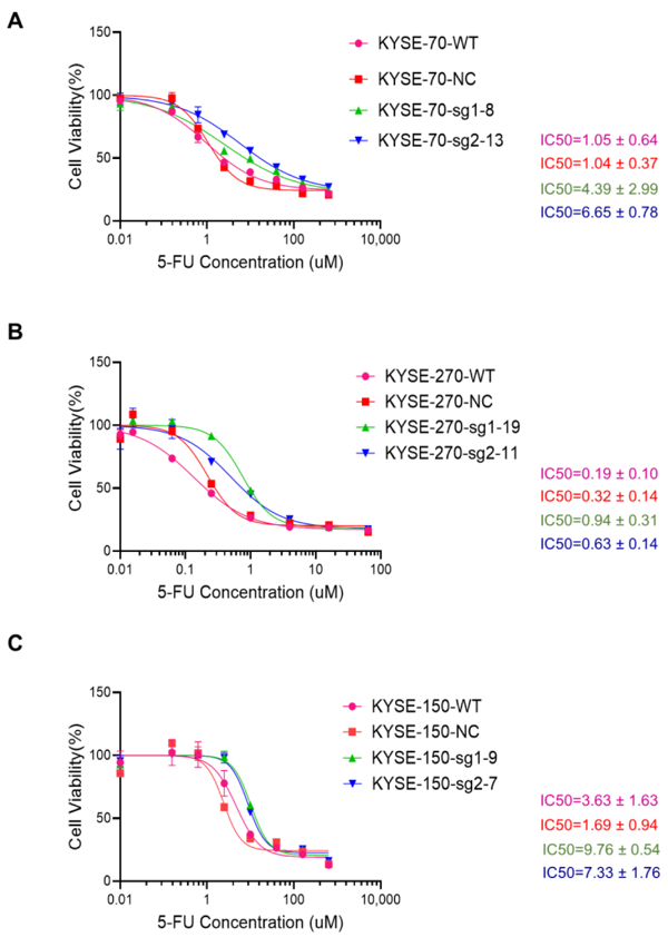
WT (wild-type cells), NC (transfected with non-targeting control sgRNA), sg1 (transfected with PFKFB3-sgRNA1), and sg2 (transfected with PFKFB3-sgRNA2).
Ask a Question
Write your own review
- You May Also Need

Description: Human moderately differentiated squamous cell carcinoma cell line established from esophageal cancer.
Description: Human moderately differentiated squamous cell carcinoma cell line established from esophageal cancer.


Description: Human moderately differentiated squamous cell carcinoma cell line established from esophageal cancer.
Description: Human squamous cell carcinoma from oral cavity via mouse transplantation.

Description: Human squamous cell carcinoma cell line established from recurrent cancer (esophageal cancer).
- Adipose Tissue-Derived Stem Cells
- Human Neurons
- Mouse Probe
- Whole Chromosome Painting Probes
- Hepatic Cells
- Renal Cells
- In Vitro ADME Kits
- Tissue Microarray
- Tissue Blocks
- Tissue Sections
- FFPE Cell Pellet
- Probe
- Centromere Probes
- Telomere Probes
- Satellite Enumeration Probes
- Subtelomere Specific Probes
- Bacterial Probes
- ISH/FISH Probes
- Exosome Isolation Kit
- Human Adult Stem Cells
- Mouse Stem Cells
- iPSCs
- Mouse Embryonic Stem Cells
- iPSC Differentiation Kits
- Mesenchymal Stem Cells
- Immortalized Human Cells
- Immortalized Murine Cells
- Cell Immortalization Kit
- Adipose Cells
- Cardiac Cells
- Dermal Cells
- Epidermal Cells
- Peripheral Blood Mononuclear Cells
- Umbilical Cord Cells
- Monkey Primary Cells
- Mouse Primary Cells
- Breast Tumor Cells
- Colorectal Tumor Cells
- Esophageal Tumor Cells
- Lung Tumor Cells
- Leukemia/Lymphoma/Myeloma Cells
- Ovarian Tumor Cells
- Pancreatic Tumor Cells
- Mouse Tumor Cells