
Ehrlich-Lettre Ascites, Strain E
Cat.No.: CSC-C9376L
Species: Mus musculus (Mouse)
Source: Ascites
Morphology: epithelial
Culture Properties: monolayer
- Specification
- Background
- Scientific Data
- Q & A
- Customer Review
Histopathology: carcinoma
Note: Tested and found negative for ectromelia virus(mousepox)
Ehrlich-Lettre Ascites, Strain E (E-La) is a classic murine tumor cell line derived from the ascitic form of an undifferentiated carcinoma first reported in Swiss albino mice. This line has been used extensively in basic cancer biology research and was derived from explant culture of in vivo passaged tumor tissue. E-La cells show an epithelial cell-like morphology under adherent culture conditions. Cytogenetically, the line is near-triploid with a modal chromosome number of about 44 (normal mouse diploid number = 40) and specific marker chromosomes, including the characteristic "A" chromosome.
For in vitro propagation, the cells require a basal medium rich in nutrients formulated for mammalian cells supplemented with serum and essential amino acids such as L-glutamine. Cultures were maintained at 37°C in humidified 5% carbon dioxide and usually sub-cultured with a standard protease solution at 70-80% confluence. The E-La strain continues to be a valuable tool for tumorigenesis studies, chemotherapeutic efficacy evaluation and modelling of peritoneal carcinomatosis because of its reliable growth characteristics and maintained malignant phenotype.
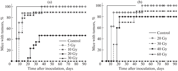
Tumor Induction Patterns in Mice Inoculated with Irradiated EAC Cells
Balakin et al. investigated the pattern of tumor induction in mice inoculated with Ehrlich ascites carcinoma (EAC) cells irradiated ex vivo with high doses of carbon ions (C) or X-rays.
As shown in Figure 1, mice inoculated with non-irradiated cells formed tumors by day 5. On the other hand, tumors appeared later in groups receiving irradiated cells in a dose-dependent fashion. The first tumors appeared on day 7 (5 Gy), day 11 (10 Gy) and day 21 (20 Gy) for C irradiation. Tumors were observed on day 11 (20 Gy), day 14 (30 Gy) and day 18 (40 Gy) after X-ray irradiation
At day 30 the tumor incidence in the C groups was 88% (5 Gy), 89% (10 Gy) and 35% (20 Gy), and no tumors were observed at 30 Gy (Fig. 1a). The incidences were 88%, 70%, 80% and 0% at 20, 30, 40 and 60 Gy, respectively, by X-ray irradiation (Fig. 1b). Tumor latency was significantly increased with dose, but the percentage of tumors induced within one month was not significantly different at lower doses for either type of radiation. This is consistent with previous studies on the proton irradiated cells, which indicate that the tumor cell death at early time points may promote the proliferation and differentiation of cancer stem cells (CSCs) in a radiation type-dependent manner.
Ask a Question
Write your own review

Description: Described as secreting a mouse monoclonal antibody (IgG2a) detecting all fibers in skeletal muscle and myosin heavy chains on Western blots and detecting mammalian, chicken, zebrafish, axolotl, ...

Description: Animals were immunized with the B6.1 mouse cytotoxic T cell line.


Description: neuroglial and neuronal character coexpressing ependymoma cell line.

Description: Established by irradiation of the adherent cells in long-term bone marrow cultures derived from C3H/HeNSlc strain mice
- Adipose Tissue-Derived Stem Cells
- Human Neurons
- Mouse Probe
- Whole Chromosome Painting Probes
- Hepatic Cells
- Renal Cells
- In Vitro ADME Kits
- Tissue Microarray
- Tissue Blocks
- Tissue Sections
- FFPE Cell Pellet
- Probe
- Centromere Probes
- Telomere Probes
- Satellite Enumeration Probes
- Subtelomere Specific Probes
- Bacterial Probes
- ISH/FISH Probes
- Exosome Isolation Kit
- Human Adult Stem Cells
- Mouse Stem Cells
- iPSCs
- Mouse Embryonic Stem Cells
- iPSC Differentiation Kits
- Mesenchymal Stem Cells
- Immortalized Human Cells
- Immortalized Murine Cells
- Cell Immortalization Kit
- Adipose Cells
- Cardiac Cells
- Dermal Cells
- Epidermal Cells
- Peripheral Blood Mononuclear Cells
- Umbilical Cord Cells
- Monkey Primary Cells
- Mouse Primary Cells
- Breast Tumor Cells
- Colorectal Tumor Cells
- Esophageal Tumor Cells
- Lung Tumor Cells
- Leukemia/Lymphoma/Myeloma Cells
- Ovarian Tumor Cells
- Pancreatic Tumor Cells
- Mouse Tumor Cells