
ESO-51
Cat.No.: CSC-C0675
Species: Homo sapiens (Human)
Source: Esophagus
Morphology: polymorphic cells growing in clusters in suspension
Culture Properties: suspension
- Specification
- Background
- Scientific Data
- Q & A
- Customer Review
Immunology: cytokeratin +, cytokeratin-7 +, cytokeratin-8 +, cytokeratin-17 -, cytokeratin-18 +, cytokeratin-19 +, desmin -, endothel -, EpCAM +, GFAP -, neurofilament -, vimentin +
Viruses: PCR:
ESO-51 is a well-characterized human cell line derived from a primary adenocarcinoma of the distal esophagus. It was established in 2000 from a 74-year-old Caucasian male, with the presence of Barrett's transformed mucosa, reflecting the metaplasia-dysplasia-adenocarcinoma sequence central to esophageal adenocarcinoma (EAC) pathogenesis. The line exhibits a polymorphic morphology, growing in large spherical clusters in suspension, and is tumorigenic in immunocompromised mice, confirming its malignant phenotype.
ESO-51 is a highly valuable model for mechanistic studies of Barrett's-associated carcinogenesis, drug sensitivity assays, and comparative genomic analyses of EAC. Its suspension growth in RPMI-1640 medium offers practical handling advantages, enabling large-scale expansion for high-throughput screening. Given the limited availability of authentic EAC models, ESO-51 serves as a crucial tool for unraveling the molecular drivers of this aggressive malignancy and for preclinical therapeutic evaluation.
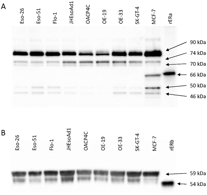
Oestrogen Receptor Isoforms May Represent a Therapeutic Target in Oesophageal Adenocarcinoma
Oesophageal adenocarcinoma is a rapidly increasing problem in which treatment options are limited. Previous studies have shown that oesophageal adenocarcinoma cells and tissues express oestrogen receptors (ERs) and show growth suppression and apoptosis in response to ER modulator agents such as tamoxifen. ERs are known to be expressed in a number of isoforms that act together to regulate cell growth and cell death.
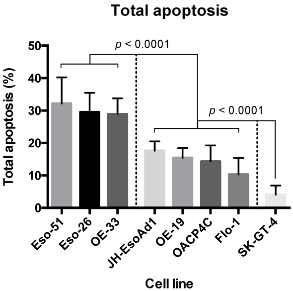
In this study, Due, Steven L., et al. used western blotting to profile the expression of ERα and ERβ isoforms, and expression of the oncologically related molecules p53, HER2, and EGFR, in a panel of oesophageal adenocarcinoma cell lines. The cytotoxicity of tamoxifen in the cell lines was determined with Annexin V-FITC flow cytometry, and correlations between cytotoxicity and receptor expression were assessed using Spearman's rank-order correlation. Oesophageal adenocarcinoma cell lines showed varying cytotoxicity in response to tamoxifen. The ER species ERα90, ERα50, and ERα46, as well as p53, were positively associated with a cytotoxic response. Conversely, ERα74, ERα70, and ERβ54 were associated with a lack of cytotoxic response. The ER species detected in oesophageal adenocarcinoma cells may work together to confer sensitivity to ER modulators in this disease, which could open up a new avenue for therapy in selected patients.
Ask a Question
Write your own review
- You May Also Need

Description: Human moderately differentiated squamous cell carcinoma cell line established from esophageal cancer.

Description: Human moderately differentiated squamous cell carcinoma cell line established from esophageal cancer.


Description: Human moderately differentiated squamous cell carcinoma cell line established from esophageal cancer.
Description: Human squamous cell carcinoma from oral cavity via mouse transplantation.

Description: Human squamous cell carcinoma cell line established from recurrent cancer (esophageal cancer).
- Adipose Tissue-Derived Stem Cells
- Human Neurons
- Mouse Probe
- Whole Chromosome Painting Probes
- Hepatic Cells
- Renal Cells
- In Vitro ADME Kits
- Tissue Microarray
- Tissue Blocks
- Tissue Sections
- FFPE Cell Pellet
- Probe
- Centromere Probes
- Telomere Probes
- Satellite Enumeration Probes
- Subtelomere Specific Probes
- Bacterial Probes
- ISH/FISH Probes
- Exosome Isolation Kit
- Human Adult Stem Cells
- Mouse Stem Cells
- iPSCs
- Mouse Embryonic Stem Cells
- iPSC Differentiation Kits
- Mesenchymal Stem Cells
- Immortalized Human Cells
- Immortalized Murine Cells
- Cell Immortalization Kit
- Adipose Cells
- Cardiac Cells
- Dermal Cells
- Epidermal Cells
- Peripheral Blood Mononuclear Cells
- Umbilical Cord Cells
- Monkey Primary Cells
- Mouse Primary Cells
- Breast Tumor Cells
- Colorectal Tumor Cells
- Esophageal Tumor Cells
- Lung Tumor Cells
- Leukemia/Lymphoma/Myeloma Cells
- Ovarian Tumor Cells
- Pancreatic Tumor Cells
- Mouse Tumor Cells