
E11(Mouse)
Cat.No.: CSC-C8794H
Species: Mus musculus (Mouse)
Source: Kidney
Morphology: Monolayer, adherent
Culture Properties: Monolayer, adherent
- Specification
- Background
- Scientific Data
- Q & A
- Customer Review
specific PCR: negative
E11 is a murine thymic lymphoma cell line, originally isolated from the thymus of a C57BL/6 mouse. It is a popular model for investigating T-cell receptor (TCR) signaling and the molecular mechanisms controlling T-cell growth, proliferation and death.
Morphologically, E11 cells are lymphoblast-like and develop in suspension as single cells or tiny aggregates. They display distinctive T cell markers such as CD4 and CD8 (double-positive phenotype) and depend on IL-2 for persistent proliferation in vitro. Under conventional conditions (37°C, 5% CO2) the cells are generally maintained in a nutrient-rich medium (e.g. RPMI-1640) supplemented with serum and exogenous IL-2.
A prominent characteristic of the E11 line is its usefulness in signal transduction studies. It is often applied to study the downstream effects of TCR ligation, calcium flux, and NF-κB activation. E11 cells are also utilized to research T-cell anergy, autophagy and immune checkpoint control making them a significant tool for immunologists studying therapeutic targets for T-cell malignancies, autoimmune diseases and transplant rejection.
Ezrin Regulates Rac1 Activity via Its N-Terminal FERM Domain in Glomerular Podocytes
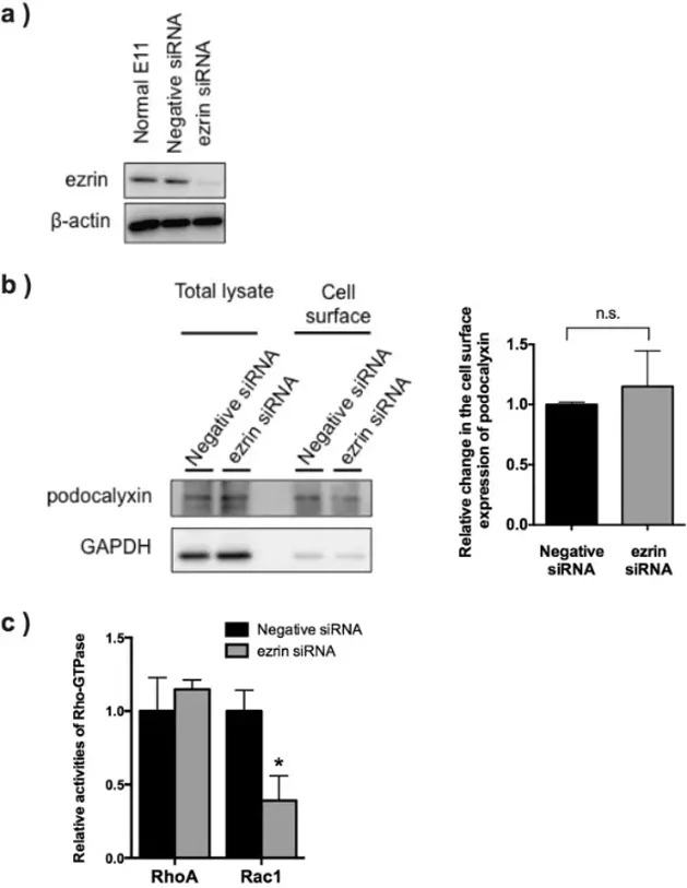
Ezrin is a key cytoskeletal protein in glomerular podocytes, forming complexes with NHERF2 and podocalyxin. While podocalyxin is essential for renal filtration, the specific physiological role of ezrin remains unclear. Hatano et al. utilized the immortalized podocyte cell line E11, which abundantly expresses ezrin (Fig. 1a), to investigate its function via siRNA-mediated knockdown.
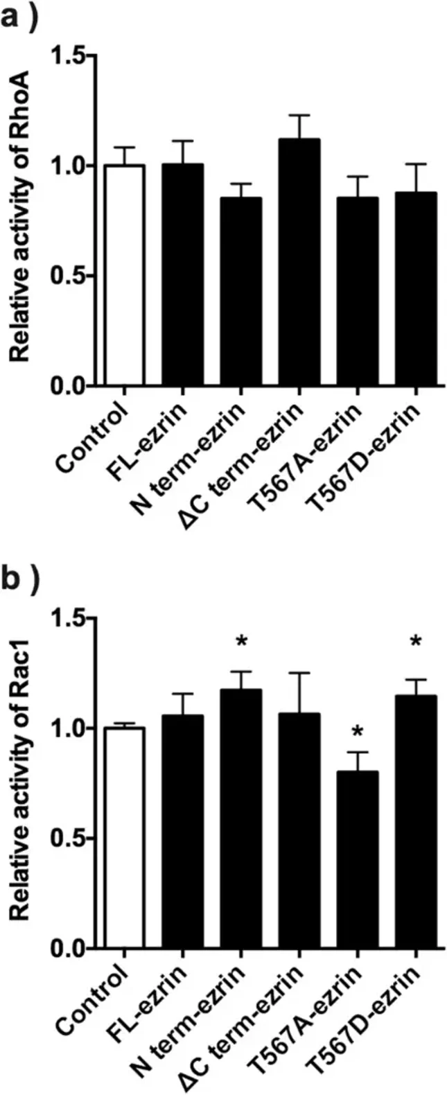
Effective ezrin depletion did not alter total podocalyxin expression (Fig. 1b, Total lysate) or its membrane localization, as confirmed by cell surface biotinylation (Fig. 1b, Cell surface). However, ezrin knockdown significantly reduced Rac1 activity without affecting RhoA activity (Fig. 1c). To dissect the structural determinants of this effect, they transfected E11 cells with various ezrin mutants. While RhoA activity remained unchanged across all constructs (Fig. 2a), Rac1 activity was significantly increased by constitutively active ezrin (T567D) and the isolated N-terminal FERM domain (N term-ezrin), and decreased by the inactive mutant (T567A) (Fig. 2b). Full-length ezrin (FL) and a C-terminal truncation lacking the actin-binding domain (ΔCt) did not significantly alter Rac1 activity (Fig. 2b). These findings indicate that ezrin regulates podocyte Rac1 activity through its N-terminal FERM domain and T567 phosphorylation, independent of podocalyxin membrane localization.
Ask a Question
Write your own review
- You May Also Need
Description: Described as secreting a mouse monoclonal antibody (IgG2a) detecting all fibers in skeletal muscle and myosin heavy chains on Western blots and detecting mammalian, chicken, zebrafish, axolotl, ...
Description: Animals were immunized with the B6.1 mouse cytotoxic T cell line.
Description: neuroglial and neuronal character coexpressing ependymoma cell line.
Description: Established by irradiation of the adherent cells in long-term bone marrow cultures derived from C3H/HeNSlc strain mice
- Adipose Tissue-Derived Stem Cells
- Human Neurons
- Mouse Probe
- Whole Chromosome Painting Probes
- Hepatic Cells
- Renal Cells
- In Vitro ADME Kits
- Tissue Microarray
- Tissue Blocks
- Tissue Sections
- FFPE Cell Pellet
- Probe
- Centromere Probes
- Telomere Probes
- Satellite Enumeration Probes
- Subtelomere Specific Probes
- Bacterial Probes
- ISH/FISH Probes
- Exosome Isolation Kit
- Human Adult Stem Cells
- Mouse Stem Cells
- iPSCs
- Mouse Embryonic Stem Cells
- iPSC Differentiation Kits
- Mesenchymal Stem Cells
- Immortalized Human Cells
- Immortalized Murine Cells
- Cell Immortalization Kit
- Adipose Cells
- Cardiac Cells
- Dermal Cells
- Epidermal Cells
- Peripheral Blood Mononuclear Cells
- Umbilical Cord Cells
- Monkey Primary Cells
- Mouse Primary Cells
- Breast Tumor Cells
- Colorectal Tumor Cells
- Esophageal Tumor Cells
- Lung Tumor Cells
- Leukemia/Lymphoma/Myeloma Cells
- Ovarian Tumor Cells
- Pancreatic Tumor Cells
- Mouse Tumor Cells