
DMBM-2
Cat.No.: CSC-C1085
Species: Mus musculus (Mouse)
Source: Bone Marrow
Morphology: round cells growing in aggregates, mostly loosely adherent, some cells have long processes
- Specification
- Background
- Scientific Data
- Q & A
- Customer Review
Viruses: ELISA: reverse transcriptase negative; PCR: SMRV -
DMBM-2 is a murine macrophage cell line derived from the bone marrow of a C3H/HeJ animal. The cell line was established by spontaneous transformation during long-term culture and frequently utilized as an in vitro model for studies on macrophage biology and innate immune responses. DMBM-2 cells have the typical macrophage morphology, characterized by round to irregular-shaped cells with mostly loose aggregation and partial adherence. Some cells exhibit lengthy cytoplasmic processes, which are indicative of their activated macrophage-like nature.
DMBM-2, as a model of bone marrow-derived macrophages, preserves many of the functional traits associated with professional phagocytes, such as response to inflammatory stimuli, generation of cytokines, and engagement in host defence mechanisms. The parental C3H/HeJ mouse strain is known for increased lipopolysaccharide (LPS) responsiveness and has been used in research of macrophage activation pathways, innate immunological signaling and mechanisms of inflammatory control, and DMBM-2 has been used in these studies.
DMBM-2 cells offer a straightforward and reproducible alternative to primary cultures of macrophages for the assessment of cytokine responses, cell-pathogen interactions and immune-modulating chemicals. They have also been employed to investigate signal transmission, macrophage differentiation, phagocytic activity and generation of inflammatory mediators. DMBM-2 cells, with their consistent growth properties and well-defined macrophage phenotype, are suitable for experimental studies in immunology, inflammation and host-pathogen interaction.
Species- and Cell-Type-Specific Lysosomal Characteristics in Macrophage Models of Phospholipidosis
Phospholipidosis (PLD), the intracellular accumulation of phospholipids, is a key toxicological response to cationic amphiphilic drugs (CADs) and can impair alveolar macrophage function. To optimize PLD screening, Öhlinger et al. evaluated a panel of lysosomal stains (LysoSensor, Acridine Orange, Nile Red, HCS LipidTOX, LysoID) across murine (DMBM-2, J774, RAW264.7) and human macrophage models (THP-1, monocyte-derived M1/M2 macrophages).
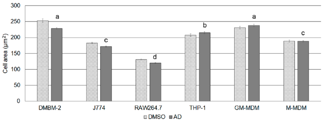
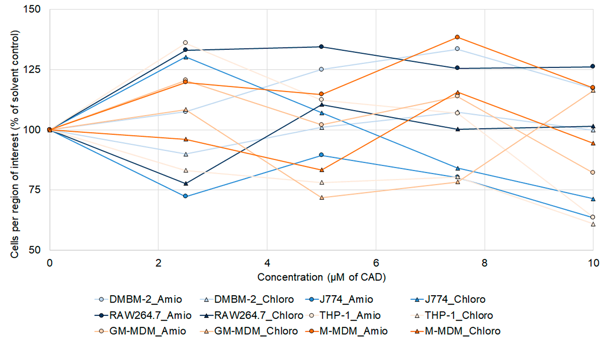
Macrophages grouped into four size categories: DMBM-2/human M1 (largest), THP-1, J774/human M2, and RAW264.7 (smallest) (Fig. 1). Lysosome quantification revealed distinct interspecies differences: murine cells had higher lysosome density (0.11-0.13/µm²) than human cells (0.05-0.06/µm²). Human macrophages possessed fewer but larger lysosomes, whereas murine macrophages had smaller, more numerous lysosomes. Correlation analysis showed strong linkage between lysosome area and dye intensity in J774 and THP-1 (r = 0.63-0.68), but weaker correlation in DMBM-2 and RAW264.7 (r = 0.42-0.48). Post-CAD exposure, cell densities varied regionally (60-134% of control) with no dose-dependency, suggesting spatial heterogeneity rather than cytotoxicity (Fig. 2). These findings highlight pronounced species-specific differences in macrophage lysosomal architecture that must be considered when selecting models for PLD assessment.
Ask a Question
Write your own review
- You May Also Need
Description: Described as secreting a mouse monoclonal antibody (IgG2a) detecting all fibers in skeletal muscle and myosin heavy chains on Western blots and detecting mammalian, chicken, zebrafish, axolotl, ...

Description: Animals were immunized with the B6.1 mouse cytotoxic T cell line.
Description: neuroglial and neuronal character coexpressing ependymoma cell line.
Description: Established by irradiation of the adherent cells in long-term bone marrow cultures derived from C3H/HeNSlc strain mice
- Adipose Tissue-Derived Stem Cells
- Human Neurons
- Mouse Probe
- Whole Chromosome Painting Probes
- Hepatic Cells
- Renal Cells
- In Vitro ADME Kits
- Tissue Microarray
- Tissue Blocks
- Tissue Sections
- FFPE Cell Pellet
- Probe
- Centromere Probes
- Telomere Probes
- Satellite Enumeration Probes
- Subtelomere Specific Probes
- Bacterial Probes
- ISH/FISH Probes
- Exosome Isolation Kit
- Human Adult Stem Cells
- Mouse Stem Cells
- iPSCs
- Mouse Embryonic Stem Cells
- iPSC Differentiation Kits
- Mesenchymal Stem Cells
- Immortalized Human Cells
- Immortalized Murine Cells
- Cell Immortalization Kit
- Adipose Cells
- Cardiac Cells
- Dermal Cells
- Epidermal Cells
- Peripheral Blood Mononuclear Cells
- Umbilical Cord Cells
- Monkey Primary Cells
- Mouse Primary Cells
- Breast Tumor Cells
- Colorectal Tumor Cells
- Esophageal Tumor Cells
- Lung Tumor Cells
- Leukemia/Lymphoma/Myeloma Cells
- Ovarian Tumor Cells
- Pancreatic Tumor Cells
- Mouse Tumor Cells