
- Specification
- Background
- Scientific Data
- Q & A
- Customer Review
The C99 cell line was derived from a moderately developed adenocarcinoma (Dukes' stage C) of the rectum of a 69-year-old male and is an important model in the field of gastrointestinal oncology. C99 is widely used in the scientific literature to study the molecular basis of chemoresistance, especially to 5-fluorouracil (5-FU) and platinum-based medicines, and the efficacy of novel targeted therapies, such as MEK and IGF-1R inhibitors.
Besides drug screening, C99 cells have greatly contributed to our knowledge on the dynamics of the tumor microenvironment. Importantly, they are characterized by low glycolytic acid generation and strong acid tolerance, making them an attractive model to investigate how cancer cells cope with acidic stress and compete metabolically in the tumor niche. C99 has also provided additional insights into the functional repercussions of E-cadherin mutations on cell adhesion, motility and invasion, especially in the context of trefoil factor signaling. These cells are maintained in regular nutritional media with serum under conventional circumstances (37°C, 5% CO2) providing a robust platform for both mechanistic research and preclinical treatment validation.
L‑Lactate Dehydrogenase B may be a Predictive Marker for Sensitivity to Anti‑EGFR Monoclonal Antibodies in Colorectal Cancer Cell Lines
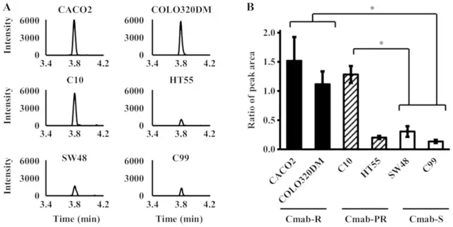
Proteins generated from cancer cells are excellent biomarkers to predict the efficacy of chemotherapy. To find indicators of anti-EGFR mAb response, Nagamine et al. analyzed proteome profiles of six CRC cell lines (CACO2, COLO320DM, C10, HT55, SW48, and C99), which show different sensitivities to cetuximab.
Mass spectrometry analyses of cytoplasmic and membrane fractions revealed 148 and 146 unique peaks for resistant lines and 363 and 267 peaks for sensitive lines, respectively. MS/MS study identified Lactate Dehydrogenase B (LDHB) as a major differential protein, especially the peptide SADTLWDIQK (m/z 1176.60). This peptide was much more intense in the cetuximab-resistant (Cmab-R) lines than in the sensitive (Cmab-S) lines (Fig. 1). Importantly, peptide C10 was significantly increased in partial resistance (Cmab-PR) compared with Cmab-S, although C99 wasn't changed a lot (Fig. 1). The MS data were linked to ELISA data.
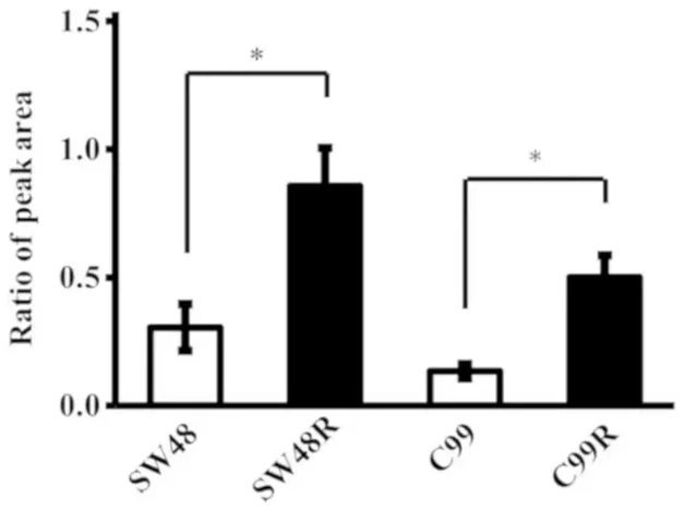
In acquired resistance models, validation of LDHB expression was dramatically elevated in cetuximab resistant derivatives (SW48R, C99R) over their parental equivalents (Fig. 2). These data suggest LDHB is a possible proteomic biomarker of cetuximab resistance in CRC.
Ask a Question
Write your own review
Description: Established from an adenocarcinoma located in the ascending colon of a 68 year-old male patient. The adenocarcinoma was well differentiated and determined to be Dukes' stage B. Imperial College ...
Description: This is one cell line out of a series of colon carcinoma cell lines established by PD Dr. Michael Linnebacher.
Description: Rectal carcinoma from a Japanese patient. Cell growth is slow.
Description: The C80 cell line was established from a 69-year old male patient with a moderately well differentiated adenocarcinoma of the rectum classified as Dukes' stage D.
Description: RKO is a poorly differentiated colon carcinoma cell line developed by Michael Brattain.
- Adipose Tissue-Derived Stem Cells
- Human Neurons
- Mouse Probe
- Whole Chromosome Painting Probes
- Hepatic Cells
- Renal Cells
- In Vitro ADME Kits
- Tissue Microarray
- Tissue Blocks
- Tissue Sections
- FFPE Cell Pellet
- Probe
- Centromere Probes
- Telomere Probes
- Satellite Enumeration Probes
- Subtelomere Specific Probes
- Bacterial Probes
- ISH/FISH Probes
- Exosome Isolation Kit
- Human Adult Stem Cells
- Mouse Stem Cells
- iPSCs
- Mouse Embryonic Stem Cells
- iPSC Differentiation Kits
- Mesenchymal Stem Cells
- Immortalized Human Cells
- Immortalized Murine Cells
- Cell Immortalization Kit
- Adipose Cells
- Cardiac Cells
- Dermal Cells
- Epidermal Cells
- Peripheral Blood Mononuclear Cells
- Umbilical Cord Cells
- Monkey Primary Cells
- Mouse Primary Cells
- Breast Tumor Cells
- Colorectal Tumor Cells
- Esophageal Tumor Cells
- Lung Tumor Cells
- Leukemia/Lymphoma/Myeloma Cells
- Ovarian Tumor Cells
- Pancreatic Tumor Cells
- Mouse Tumor Cells