B9
Cat.No.: CSC-C6191X
Species: Mus musculus (Mouse)
Morphology: round cells growing in suspension
Culture Properties: Suspension
- Specification
- Background
- Scientific Data
- Q & A
- Customer Review
B9 cell line is a well-characterized murine hybridoma cell line widely used as a classical bioassay model for detecting and quantifying interleukin-6 (IL-6) biological activity. Originally derived from murine B-cell lineage hybridoma technology, B9 cells are strictly IL-6-dependent for proliferation, making them highly sensitive and specific indicators of IL-6 signaling. In the absence of IL-6, B9 cells do not proliferate, while exposure to IL-6 or IL-6-containing samples induces robust cell growth, which can be quantified using colorimetric or proliferation-based assays.
Because of this unique dependency, B9 cells have become a gold-standard functional assay system in immunology and inflammation research. They are extensively used to measure IL-6 levels in biological samples, evaluate cytokine secretion in disease models, and assess the neutralizing activity of anti-IL-6 or anti-IL-6 receptor antibodies. In addition, B9-based assays are frequently applied in studies of autoimmune diseases, cancer-related inflammation, infection-induced cytokine storms, and therapeutic antibody development.
Although modern ELISA and multiplex platforms have supplemented cytokine detection technologies, B9 cells remain valuable for functional bioactivity assessment, as they reflect the biological rather than merely the quantitative presence of IL-6. Their high sensitivity and reproducibility continue to support both basic research and translational studies targeting IL-6-mediated signaling pathways.
Quantitative Analysis of IL-6 Binding and Bioactivity in B9 Cells
Despite the clinical importance of cytokines like IL-6, the quantitative relationship between molecular binding and cellular activation thresholds remains unclear. Utilizing validated tools such as iodinated IL-6 and B9 hybridoma bioassays, Hansen et al. employed equilibrium binding principles and experimental data to estimate cellular IL-6 interactions in blood.
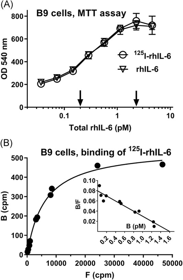
They characterized IL-6 binding in the IL-6-dependent B9 hybridoma cell line using 125I-labeled rhIL-6 (Fig. 1A). Bioactivity, measured by cell viability, was detectable at a total IL-6 concentration of 0.2 pM and maximized at 2.3 pM (Fig. 1A). Scatchard analysis revealed approximately 164 high-affinity binding sites per cell (Kd ≈ 20 pM) (Fig. 1B).
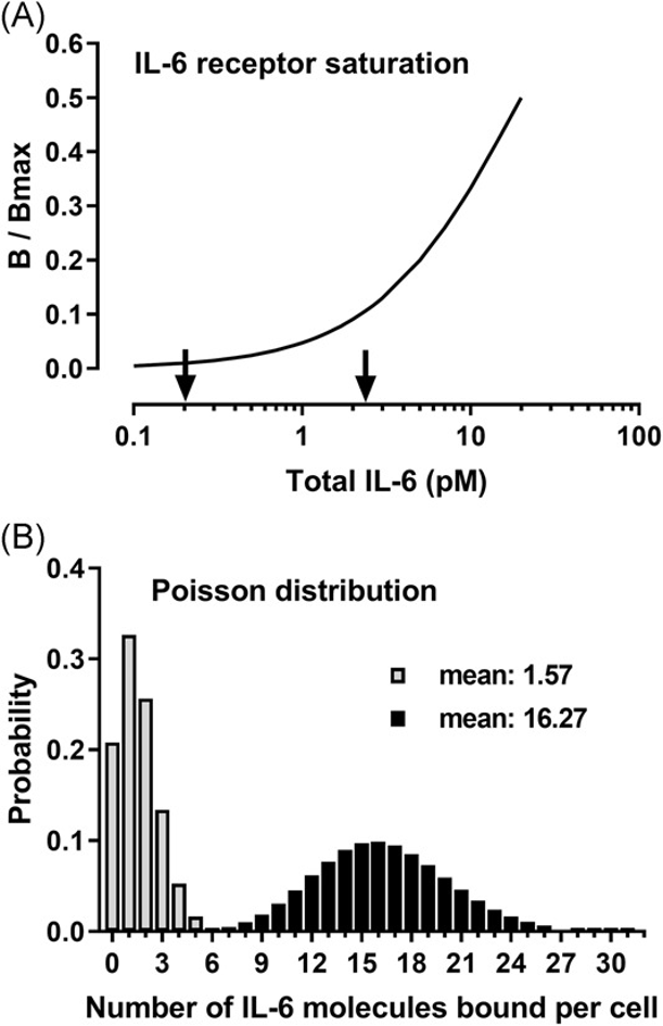
At the threshold bioactive concentration (0.2 pM total IL-6), cells bound an average of 1.57 molecules, corresponding to just 1% receptor occupancy (Fig. 2A). This minimal binding was sufficient to trigger a measurable proliferative signal. Poisson distribution analysis indicated that at this concentration, 46.5% of cells bound ≥2 IL-6 molecules (Fig. 2B).
At the maximal bioactivity concentration (2.3 pM), cells bound an average of 16.27 molecules. Given that the IL-6 receptor complex functions as a hexamer requiring two IL-6 molecules, these data suggest that activation of merely two receptors (four IL-6 molecules) is sufficient for measurable bioactivity, while activation of approximately eight receptors drives maximal response in vitro.
Ask a Question
Write your own review
- You May Also Need

Description: Described as secreting a mouse monoclonal antibody (IgG2a) detecting all fibers in skeletal muscle and myosin heavy chains on Western blots and detecting mammalian, chicken, zebrafish, axolotl, ...

Description: Animals were immunized with the B6.1 mouse cytotoxic T cell line.


Description: neuroglial and neuronal character coexpressing ependymoma cell line.

Description: Established by irradiation of the adherent cells in long-term bone marrow cultures derived from C3H/HeNSlc strain mice
- Adipose Tissue-Derived Stem Cells
- Human Neurons
- Mouse Probe
- Whole Chromosome Painting Probes
- Hepatic Cells
- Renal Cells
- In Vitro ADME Kits
- Tissue Microarray
- Tissue Blocks
- Tissue Sections
- FFPE Cell Pellet
- Probe
- Centromere Probes
- Telomere Probes
- Satellite Enumeration Probes
- Subtelomere Specific Probes
- Bacterial Probes
- ISH/FISH Probes
- Exosome Isolation Kit
- Human Adult Stem Cells
- Mouse Stem Cells
- iPSCs
- Mouse Embryonic Stem Cells
- iPSC Differentiation Kits
- Mesenchymal Stem Cells
- Immortalized Human Cells
- Immortalized Murine Cells
- Cell Immortalization Kit
- Adipose Cells
- Cardiac Cells
- Dermal Cells
- Epidermal Cells
- Peripheral Blood Mononuclear Cells
- Umbilical Cord Cells
- Monkey Primary Cells
- Mouse Primary Cells
- Breast Tumor Cells
- Colorectal Tumor Cells
- Esophageal Tumor Cells
- Lung Tumor Cells
- Leukemia/Lymphoma/Myeloma Cells
- Ovarian Tumor Cells
- Pancreatic Tumor Cells
- Mouse Tumor Cells