
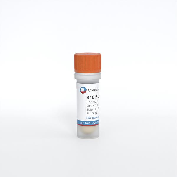
B16 BL6
Cat.No.: CSC-C9334L
Species: Mus musculus (Mouse)
Source: Skin
Culture Properties: monolayer
- Specification
- Background
- Scientific Data
- Q & A
- Customer Review
B16-BL6 is a well-characterized murine melanoma cell line derived as a metastatic subline of the B16 melanoma, which was originally established in 1954 from a spontaneous skin tumor in a C57BL/6 mouse. The B16-BL6 subline was specifically selected from the B16-F10 parental line for enhanced tissue-invasive properties, exhibiting greater metastatic capacity than its predecessors. The cells display a spindle- to polygonal-shaped morphology, adhere in monolayers, and remain pigmented due to tyrosinase expression. Genetically, B16-BL6 carries TP53 mutations and displays severe chromosomal heteroploidy, while lacking BRAF, NRAS, and PTEN mutations-a mutational profile distinct from human melanoma.
The defining advantage of B16-BL6 lies in its exceptional metastatic potential, particularly to the lungs following intravenous injection. This feature, combined with its syngeneic compatibility with C57BL/6 mice, enables immune-competent studies of tumor progression, metastatic dissemination, and tumor-immune interactions. The cells exhibit low MHC class I/II expression, contributing to immune evasion and making B16-BL6 an ideal platform for evaluating immunotherapies and checkpoint inhibitors. B16-BL6 also demonstrates elevated MSH/ACTH-responsive adenylate cyclase activity, linking hormonal signaling to metastatic behavior. Upon subcutaneous implantation, the cells form aggressive, rapidly growing tumors within 7-10 days.
Anti-Hyperpigmentation-Related Potential Activities of Essential Oil from Chamaecyparis pisifera Leaves in B16BL6 Cells
Chamaecyparis pisifera (C. pisifera; family Cupressaceae) is known to have insecticidal and antibacterial activities, but its effects on skin depigmentation-related activities have not been elucidated. Thus, in the present study, we aimed to investigate the anti-hyperpigmentation potential of C. pisifera var. filifera leaf essential oil (CPEO), specially focusing on responses related to melanogenesis and melanin transport, using B16BL6 cells. The biological activities of CPEO on B16BL6 melanoma cells were analyzed using the water-soluble tetrazolium salt, BrdU incorporation, ELISA, and immunoblotting assays.
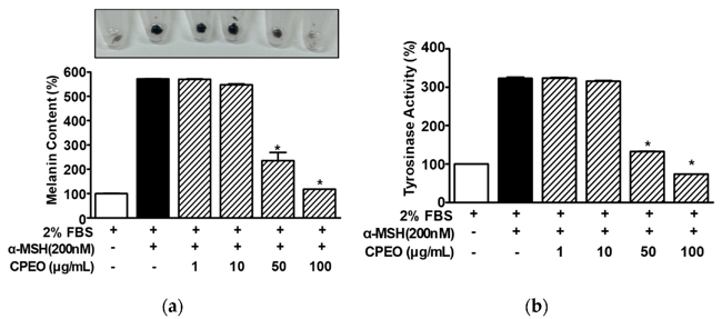
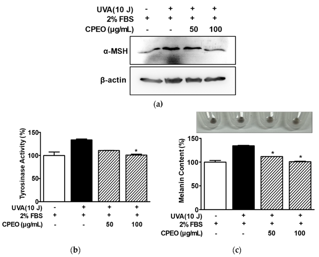
CPEO was noncytotoxic to B16BL6 cells at 1-100 μg/mL and reduced serum-induced proliferation in B16BL6 cells. CPEO significantly inhibited α-MSH-stimulated increases in melanin synthesis and tyrosinase activity in the cells. CPEO also downregulated the expression levels of melanogenesis-related proteins (MITF, tyrosinase, TRP-1 and -2) and melanosome transport-related proteins (Rab27a, melanophilin, myosin Va) in cells exposed to α-MSH. Moreover, the essential oil increased the phosphorylations of MAPKs (p38, ERK1/2, and JNK) in α-MSH-treated B16BL6 cells. In addition, conditioned medium from HaCaT cells irradiated with UVA (CM-UVA) in the presence of CPEO reduced melanin synthesis and tyrosinase activity in B16BL6 cells. These findings suggest that CPEO inhibits melanin production (probably through the regulation of MAPKs) and transport-related activities in B16BL6 cells, and that CPEO may serve as a potential natural anti-hyperpigmentation or skin whitening.
Ask a Question
Write your own review
- You May Also Need
Description: Described as secreting a mouse monoclonal antibody (IgG2a) detecting all fibers in skeletal muscle and myosin heavy chains on Western blots and detecting mammalian, chicken, zebrafish, axolotl, ...
Description: Animals were immunized with the B6.1 mouse cytotoxic T cell line.
Description: neuroglial and neuronal character coexpressing ependymoma cell line.
Description: Established by irradiation of the adherent cells in long-term bone marrow cultures derived from C3H/HeNSlc strain mice
- Adipose Tissue-Derived Stem Cells
- Human Neurons
- Mouse Probe
- Whole Chromosome Painting Probes
- Hepatic Cells
- Renal Cells
- In Vitro ADME Kits
- Tissue Microarray
- Tissue Blocks
- Tissue Sections
- FFPE Cell Pellet
- Probe
- Centromere Probes
- Telomere Probes
- Satellite Enumeration Probes
- Subtelomere Specific Probes
- Bacterial Probes
- ISH/FISH Probes
- Exosome Isolation Kit
- Human Adult Stem Cells
- Mouse Stem Cells
- iPSCs
- Mouse Embryonic Stem Cells
- iPSC Differentiation Kits
- Mesenchymal Stem Cells
- Immortalized Human Cells
- Immortalized Murine Cells
- Cell Immortalization Kit
- Adipose Cells
- Cardiac Cells
- Dermal Cells
- Epidermal Cells
- Peripheral Blood Mononuclear Cells
- Umbilical Cord Cells
- Monkey Primary Cells
- Mouse Primary Cells
- Breast Tumor Cells
- Colorectal Tumor Cells
- Esophageal Tumor Cells
- Lung Tumor Cells
- Leukemia/Lymphoma/Myeloma Cells
- Ovarian Tumor Cells
- Pancreatic Tumor Cells
- Mouse Tumor Cells