Rabbit Adipose-Derived Mesenchymal Stem Cells
- Specification
- Background
- Scientific Data
- Q & A
- Customer Review
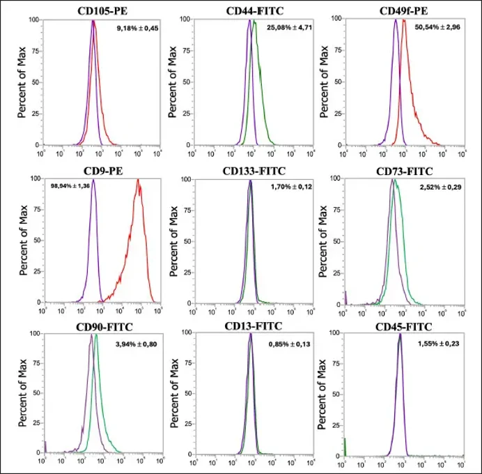
Rabbit adipose-derived mesenchymal stem cells (rAD-MSCs) are multipotent stromal cells isolated from the adipose tissue of rabbits. These cells exhibit characteristic fibroblast-like spindle-shaped morphology, robust plastic adherence, and a population doubling time consistent with active proliferation in culture. rAD-MSCs express a panel of mesenchymal stem cell surface markers including CD29, CD44, CD73, CD90, and CD105, while remaining negative for the hematopoietic markers CD34 and CD45. Additionally, they express pluripotency-associated transcription factors such as NANOG, OCT4, and SOX2. Notably, CD81 has been proposed as a stable alternative marker for rabbit MSC characterization.
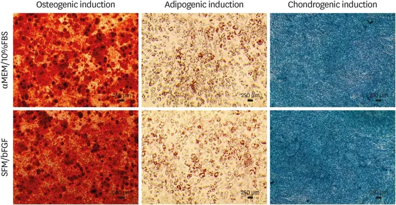
A defining advantage of rAD-MSCs is their robust trilineage differentiation potential, with demonstrated capacity to differentiate into osteocytes, adipocytes, and chondrocytes under appropriate induction conditions. They also possess neurogenic differentiation capability, as evidenced by expression of neuronal markers ENO2 and MAP2. Compared to human adipose-derived MSCs, rAD-MSCs exhibit greater clonogenic potential and proliferation rates, making them particularly advantageous for large-scale expansion.
Basic Fibroblast Growth Factor Preserves Rabbit Adipose-Derived Mesenchymal Stromal Cells in Serum-Free Culture
To develop and evaluate a rabbit adipose-derived mesenchymal stromal cell (Rab-ADMSC) serum-free medium (SFM) supplemented with growth factors that maintains key MSC characteristics.
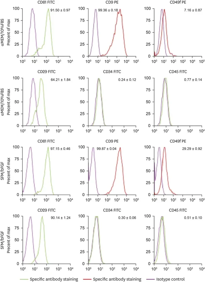
Rab-ADMSCs were isolated from 3 male New Zealand White rabbits. Basic fibroblast growth factor (bFGF), transforming growth factor-β1, and insulin-like growth factor-1 were screened for support of cell viability in SFM. Cells were then cultured in α-Minimum Essential Medium with 10% FBS or in SFM supplemented with bFGF and assessed for morphology, proliferation, expression of stemness- and senescence-associated transcripts, surface marker expression, and tri-lineage differentiation potential.
SFM supplemented with bFGF preserved fibroblast-like morphology and supported Rab-ADMSC expansion. Transcript levels of stemness-associated markers and senescence-associated markers were comparable between culture conditions. Rab-ADMSCs retained osteogenic, adipogenic, and chondrogenic differentiation potential. Flow cytometry showed higher expression of CD81, CD49f, and CD29 in the SFM plus bFGF condition, whereas CD34 and CD45 remained low in both conditions.
Ask a Question
Write your own review
- Adipose Tissue-Derived Stem Cells
- Human Neurons
- Mouse Probe
- Whole Chromosome Painting Probes
- Hepatic Cells
- Renal Cells
- In Vitro ADME Kits
- Tissue Microarray
- Tissue Blocks
- Tissue Sections
- FFPE Cell Pellet
- Probe
- Centromere Probes
- Telomere Probes
- Satellite Enumeration Probes
- Subtelomere Specific Probes
- Bacterial Probes
- ISH/FISH Probes
- Exosome Isolation Kit
- Human Adult Stem Cells
- Mouse Stem Cells
- iPSCs
- Mouse Embryonic Stem Cells
- iPSC Differentiation Kits
- Mesenchymal Stem Cells
- Immortalized Human Cells
- Immortalized Murine Cells
- Cell Immortalization Kit
- Adipose Cells
- Cardiac Cells
- Dermal Cells
- Epidermal Cells
- Peripheral Blood Mononuclear Cells
- Umbilical Cord Cells
- Monkey Primary Cells
- Mouse Primary Cells
- Breast Tumor Cells
- Colorectal Tumor Cells
- Esophageal Tumor Cells
- Lung Tumor Cells
- Leukemia/Lymphoma/Myeloma Cells
- Ovarian Tumor Cells
- Pancreatic Tumor Cells
- Mouse Tumor Cells