
Immortalized Mouse Cardiac Fibroblasts-SV40
Cat.No.: CSC-I2177Z
Species: mouse
Morphology: Polygonal
Culture Properties: Adherent
- Specification
- Background
- Scientific Data
- Q & A
- Customer Review
free from contaminations (bacteria incl. mycoplasma, fungi, HIV, HAV, HBV, HCV, Parvo-B19) and cross-contaminations
Note: Never can cells be kept at -20°C.
Immortalized Mouse Cardiac Fibroblasts-SV40 is a mouse cell line that originated from primary cardiac fibroblasts and then were immortalized with SV40 large T antigen. They have been shown to proliferate indefinitely in culture while retaining several characteristics specific to cardiac fibroblasts. These cells provide a reliable and reproducible platform to study cardiac fibroblast function in vitro.
Like normal cardiac fibroblasts, SV40-immortalized mouse cardiac fibroblasts grow as adherent cells and have a fibroblast-like spindle shape. They express markers specific to fibroblasts including vimentin, fibroblast-specific protein 1 (FSP1), α-smooth muscle actin (α-SMA) and type I collagen (COL1A1). Fibroblasts are responsible for synthesis and remodeling of the extracellular matrix (ECM) and expression of these proteins demonstrates that these cells continue to function as fibroblasts would in vivo. These cells can also proliferate, migrate, produce cytokines, and are able to differentiate into myofibroblasts upon stimulation.
These cells have been used as a tool to study cardiac fibrosis, cardiac remodeling that occurs after injury to the heart, and the molecular signaling involved in activation of cardiac fibroblasts. Researchers can leverage these cells to explore the signaling pathways involved in fibroblast processes like proliferation, myofibroblast transformation, and ECM formation, such as TGF-β/Smad, NF-κB, MAPK, and Wnt/β-catenin signaling. They can also be used for drug screening, testing the efficacy of anti-fibrotic drugs, and studying the mechanisms of cardiac remodeling.
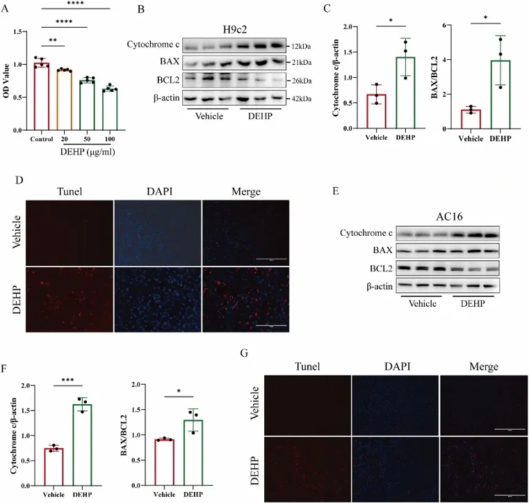
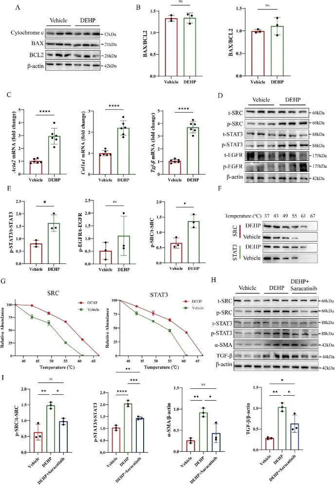
DEHP Promotes Apoptosis of Cardiomyocytes and the Transformation of Cardiac Fibroblasts into Myofibroblasts
Di-(2-ethylhexyl) phthalate (DEHP) has been epidemiologically associated with cardiac fibrosis. However, its underlying molecular mechanism remains elusive. Here, Zhang's team performed network toxicology analyses combined with molecular dynamics simulations and in vitro experiments to understand the cardiotoxic mechanism of DEHP.
Treatment with DEHP decreased cell viability of cardiomyocytes in a dose-dependent manner determined by CCK-8 assays, and thus 50 μg/mL DEHP was used for the following studies (Fig. 1A). Treatment with 50 μg/mL DEHP for 24 h significantly increased TUNEL-positive cardiomyocytes in rat H9c2 and human AC16 cells (Fig. 1D, G) and increased the ratio of BAX to BCL2 as well as cytochrome c expression in these cells (Fig. 1B, C, E, F), which demonstrated that DEHP induced apoptosis in cardiomyocytes. Interestingly, treatment with 50 μg/mL DEHP for 24 h did not induce apoptosis in immortalized mouse cardiac fibroblasts (Fig. 2A, B) but significantly increased fibrosis-associated markers including Tgfb1, Col1a1, and Acta2, markers of myofibroblast transdifferentiation (Fig. 2C). Mechanistically, they postulated that DEHP may exacerbate cardiac dysfunction via SRC, STAT3, and EGFR signaling axis. Western blot analyses revealed that the total protein level of SRC, STAT3, or EGFR were not changed upon DEHP treatment but p-SRC and p-STAT3 protein levels were significantly increased with no change in p-EGFR protein level, which confirmed the selective activation of SRC-STAT3 signaling axis (Fig. 2D, E). They performed CETSA and found that DEHP bound to and thermally stabilized SRC and STAT3 (Fig. 2F, G). Further, saracatinib (5 μM), an SRC/STAT3 phosphorylation inhibitor, rescued DEHP-induced increase in TGF-β and ACTA2 expression (Fig. 2H, I). Taken together, they uncovered that DEHP induced cardiomyocyte apoptosis and promoted cardiac fibrosis through direct activation of SRC-STAT3 signaling.
Ask a Question
Write your own review

Description: Immortalized Cynomolgus Monkey Pulmonary Artery Smooth Muscle Cells-GFP provided by Creative Bioarray have been developed by immortalizing cynomolgus monkey pulmonary artery smooth muscle cells with ...
Description: Immortalized Monkey Spleen Fibroblasts-SV40T were developed from monkey tissues transduced with a lentiviral expression vector containing the SV40T gene. The cell line was continuously cultured for ...
Description: Immortalized Monkey Bronchial Fibroblasts-GFP provided by Creative Bioarray have been developed by immortalizing monkey bronchial fibroblasts with SV40 Large T antigen and transfecting with tGFP. The ...
Description: Immortalized Canine Kidney Fibroblasts-SV40 have been obtained immortalizing Canine Kidney Fibroblasts with Lenti-SV40 Lentivirus. Immortalized cells were controlled passaging side by side with the ...
Description: Immortalized Monkey Primary Aortic Endothelial Cells-GFP provided by Creative Bioarray have been developed by immortalizing primary monkey aortic endothelial cells with SV40 Large T antigen and ...
Description: Immortalized Monkey Pancreatic Epithelial Cells-SV40T were developed from monkey tissues transduced with a lentiviral expression vector containing the SV40T gene. The cell line was continuously ...
- Adipose Tissue-Derived Stem Cells
- Human Neurons
- Mouse Probe
- Whole Chromosome Painting Probes
- Hepatic Cells
- Renal Cells
- In Vitro ADME Kits
- Tissue Microarray
- Tissue Blocks
- Tissue Sections
- FFPE Cell Pellet
- Probe
- Centromere Probes
- Telomere Probes
- Satellite Enumeration Probes
- Subtelomere Specific Probes
- Bacterial Probes
- ISH/FISH Probes
- Exosome Isolation Kit
- Human Adult Stem Cells
- Mouse Stem Cells
- iPSCs
- Mouse Embryonic Stem Cells
- iPSC Differentiation Kits
- Mesenchymal Stem Cells
- Immortalized Human Cells
- Immortalized Murine Cells
- Cell Immortalization Kit
- Adipose Cells
- Cardiac Cells
- Dermal Cells
- Epidermal Cells
- Peripheral Blood Mononuclear Cells
- Umbilical Cord Cells
- Monkey Primary Cells
- Mouse Primary Cells
- Breast Tumor Cells
- Colorectal Tumor Cells
- Esophageal Tumor Cells
- Lung Tumor Cells
- Leukemia/Lymphoma/Myeloma Cells
- Ovarian Tumor Cells
- Pancreatic Tumor Cells
- Mouse Tumor Cells