
Immortalized Equine Kidney Cells (extEqFK)
Cat.No.: CSC-I9292L
Species: Equine
Source: Kidney
Morphology: Polygonal
Culture Properties: Adherent
- Specification
- Background
- Scientific Data
- Q & A
- Customer Review
Note: Never can cells be kept at -20 °C.
2) PCR analysis for HPV16 E6/E7 presence
Immortalized Equine Kidney Cells (extEqFK) are an established cell line from the kidney of the horse (Equus caballus) originally created for use in research to allow for long-term and renewable cultures of equine kidney cells in vitro. Immortalization of extEqFK cells was achieved by transduction with human papillomavirus 16 (HPV16) E6/E7 oncogenes through non-replicating retroviral transduction (PA317 LXSN HPV16 E6/E7).
The extEqFK cell line is adherent with epithelial like morphology growing as a monolayer in vitro, like their tissue of origin. These cells are commonly used to model renal cell biology, host-pathogen interactions, and equine-specific cellular responses. Applications of extEqFK primarily include their use in veterinary virology research where they are commonly utilized to study equine infections. ExtEqFK cells have also been used in toxicology/pharmacology assays as well as research into kidney-specific cell biology from the horse. Immortalized Equine Kidney cells can be passaged indefinitely in culture, overcoming the problems of primary cells while maintaining phenotypic and functional consistency.
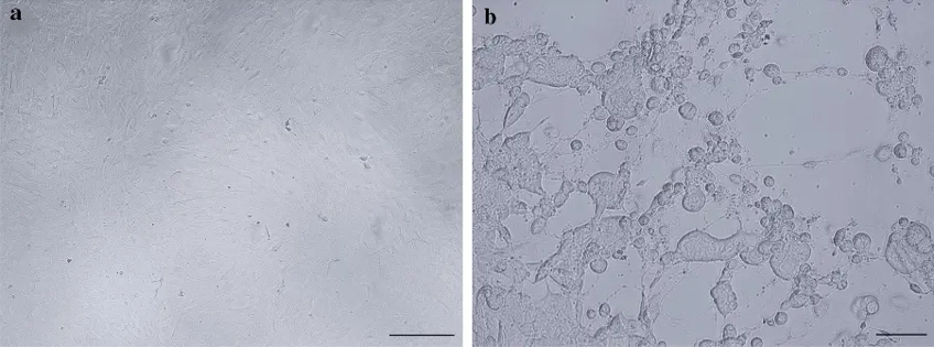
Isolation of Equid Alphaherpesvirus 3 from a Horse in Iceland with Equine Coital Exanthema
Equine coital exanthema (ECE) caused by equid alphaherpesvirus 3 (EHV-3) is a contagious venereal disease. Three types of equine herpesviruses (EHV) have been found in Iceland, EHV-4, EHV-2 and EHV-5, while EHV-1 has never been detected. Thorsteinsdóttir et al. isolated EHV-3 from a mare with papules on the vulva and inoculated in equine kidney cells (Fig. 1). It has been shown that EHV-3 replicates almost exclusively in cells of equine origin. They therefore tested the growth of dilutions of the EHV-3 virus stock in five different cells culture systems and determined the cytopathy with a light microscope. Three different equine fetal cells were tested, both primary cells and cells with extended life span. The primary cells can be passage about 10-12 times as compared to 40 passages for the cells with extended life span. The titers were 105.7, 105.11 and 105.56 TCID50/mL, respectively, for the three equine cell culture tested, prmEqFK, extEqFK and extEqFL. As expected, no CPE was observed in the RK13 and Vero cells as they are not equine cells.
Ask a Question
Write your own review

Description: Immortalized Cynomolgus Monkey Pulmonary Artery Smooth Muscle Cells-GFP provided by Creative Bioarray have been developed by immortalizing cynomolgus monkey pulmonary artery smooth muscle cells with ...
Description: Immortalized Monkey Spleen Fibroblasts-SV40T were developed from monkey tissues transduced with a lentiviral expression vector containing the SV40T gene. The cell line was continuously cultured for ...
Description: Immortalized Monkey Bronchial Fibroblasts-GFP provided by Creative Bioarray have been developed by immortalizing monkey bronchial fibroblasts with SV40 Large T antigen and transfecting with tGFP. The ...

Description: Immortalized Canine Kidney Fibroblasts-SV40 have been obtained immortalizing Canine Kidney Fibroblasts with Lenti-SV40 Lentivirus. Immortalized cells were controlled passaging side by side with the ...
Description: Immortalized Monkey Primary Aortic Endothelial Cells-GFP provided by Creative Bioarray have been developed by immortalizing primary monkey aortic endothelial cells with SV40 Large T antigen and ...
Description: Immortalized Monkey Pancreatic Epithelial Cells-SV40T were developed from monkey tissues transduced with a lentiviral expression vector containing the SV40T gene. The cell line was continuously ...
- Adipose Tissue-Derived Stem Cells
- Human Neurons
- Mouse Probe
- Whole Chromosome Painting Probes
- Hepatic Cells
- Renal Cells
- In Vitro ADME Kits
- Tissue Microarray
- Tissue Blocks
- Tissue Sections
- FFPE Cell Pellet
- Probe
- Centromere Probes
- Telomere Probes
- Satellite Enumeration Probes
- Subtelomere Specific Probes
- Bacterial Probes
- ISH/FISH Probes
- Exosome Isolation Kit
- Human Adult Stem Cells
- Mouse Stem Cells
- iPSCs
- Mouse Embryonic Stem Cells
- iPSC Differentiation Kits
- Mesenchymal Stem Cells
- Immortalized Human Cells
- Immortalized Murine Cells
- Cell Immortalization Kit
- Adipose Cells
- Cardiac Cells
- Dermal Cells
- Epidermal Cells
- Peripheral Blood Mononuclear Cells
- Umbilical Cord Cells
- Monkey Primary Cells
- Mouse Primary Cells
- Breast Tumor Cells
- Colorectal Tumor Cells
- Esophageal Tumor Cells
- Lung Tumor Cells
- Leukemia/Lymphoma/Myeloma Cells
- Ovarian Tumor Cells
- Pancreatic Tumor Cells
- Mouse Tumor Cells