
Renal Medullary Epithelial Cells, Primary, 500,000 cells per vial
Cat.No.: CSC-C4069X
Species: Human
Source: Kidney
Cell Type: Epithelial Cell
- Specification
- Background
- Scientific Data
- Q & A
- Customer Review
Cell Features:
Mixed Renal Epithelial, Renal Cortical Epithelial, and Renal Medullary Epithelial are cryopreserved as primary cells isolated from human kidney tissue and expanded in culture vessels once before cryopreservation.
Renal Proximal Tubule Epithelial are cryopreserved as secondary cells isolated from human kidney tissue and expanded in culture vessels twice before cryopreservation.
All Renal Epithelial Cell types can be grown in a 0.5% serum medium without phenol red or antimicrobials when cultured in LIRen Medium.
All Renal Epithelial Cell types are extensively tested for quality and optimal performance.
Creative Bioarray guarantees performance and quality.
Human renal medullary epithelial cells are epithelial cells isolated from the renal medulla of the human kidney. They are often isolated from the collecting ducts and the medullary thick ascending limb (mTAL) of Henle's loop. Renal medullary epithelial cells play an important role in water and electrolyte homeostasis. They are also specialized for urine concentrating mechanisms operating under physiologically hyperosmotic conditions.
The cells grow adherently and demonstrate epithelial cobblestone morphology in culture and express epithelial markers and markers specific to renal tubular epithelium such as E-cadherin, cytokeratins, aquaporins (AQP2 is often found in collecting duct derived cells), and uromodulin (found in medullary thick ascending limb derived cells). Human renal medullary epithelial cells also share functionality with their in vivo counterparts such as ion transport activity, water permeability, and hormone responsiveness. These cells also have high tolerance to the physiologically hyperosmotic microenvironment found in the renal medulla caused by high concentrations of sodium, urea and other solutes. Cells isolated from this region activate cellular mechanisms for protection against hyperosmotic conditions such as accumulating osmolytes, stress response signaling pathways (like NFAT5/TonEBP), and antioxidant responses.
Renal medullary epithelial cells serve as an in vitro model to study renal physiology and pathophysiology including: renal osmoregulation, ion transport, acute kidney injury (AKI), chronic kidney disease (CKD), tubular dysfunction, inflammation and fibrosis. They are also used to study drug-induced kidney damage and renal pharmacology.
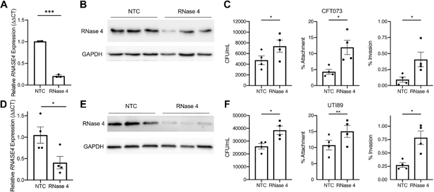
Silencing RNase 4 Expression Increases UPEC Susceptibility
Antimicrobial peptides are critical host defenses against urinary tract infections (UTIs). Ribonuclease A superfamily peptides show antimicrobial activity against uropathogens, but RNase 4's role in the human urinary tract remains unexplored. Bender et al. investigated RNase 4's antibacterial function, tissue expression, and contribution to UTI defense.
To confirm that suppressed RNase 4 expression potentiates invasive UPEC infection, RNase 4 was silenced in primary human renal medullary epithelial cells and bladder urothelial cells using siRNA. Quantitative RT-PCR and Western blot confirmed efficient knockdown in both cell types (Fig. 1A, B, D, and E), with no effect on RNASE5 expression. ELISA verified reduced RNase 4 secretion into culture media, and antibody specificity was confirmed by blocking experiments and lack of cross-reactivity with recombinant RNase 5.
Culture media collected from transfected cells were inoculated with UPEC. After 90 min, bacterial survival significantly increased in media from RNASE4-silenced cells (Fig. 1C and F). In direct infection assays, RNase 4 knockdown significantly enhanced UPEC binding and invasion in both kidney and bladder cells (Fig. 1C and F), with similar results observed in human 5,637 urothelial cells. These findings demonstrate that RNase 4 protects the kidney and bladder uroepithelium from UPEC infection.
Ask a Question
Write your own review

Description: Renal glomerular endothelial cells (GEC) are a specialized microvascular cell type involved in the regulation of glomerular ultrafiltration. They form the inner part of the filtration barrier and are ...

Description: Human Kidney Endothelial Cells from Creative Bioarray are isolated from human kidney tissue. Human Kidney Endothelial Cells are grown in T25 tissue culture flasks pre-coated with gelatin-based ...
Description: Human Proximal Tubular Epithelial Cells from Creative Bioarray are isolated from normal human proximal tubular tissue. Human Proximal Tubular Epithelial Cells are grown in T25 tissue culture flasks ...

Description: Primary Human Glomerular Microvascular Endothelial Cells were initiated by decapsulated glomeruli isolated from normal human kidney cortical tissue.These cells were originated using Complete ...
Description: Human Kidney Carcinoma Epithelial Cells from Creative Bioarray are isolated from human kidney tumor tissue. Human Kidney Carcinoma Epithelial Cells are grown in T25 tissue culture flasks pre-coated ...
Description: VE-Cad-GFP Expressing Human Glomerular Microvascular Endothelial Cells (VE-Cad-GFP HGMvECs) provided by Creative Bioarray are puromycin-selected from Human Glomerular Microvascular Endothelial Cells ...