Annexin V Apoptosis Assay
As a new method for detecting apoptosis, the Annexin V binding assay is based on the measurement of the loss of plasma membrane asymmetry. Under normal physiological conditions, cell sustain a strictly asymmetric distribution of phospholipids in the two leaflets of the cellular membranes, where phosphatidylserine (PS) facing the cytosolic side. In the early stages of apoptosis, this asymmetry disappeared rapidly, while the integrity of the membrane remained, which resulted in the exposure of PS to the outer leaflet by means of phagocytosis. This phenomenon can be detected by hapten-labeled Annexin V, which shows high affinity for PS residues in the presence of micromolar concentrations of Ca2+. By simultaneously excluding nuclear dye propidium iodide (PI), apoptotic cells and necrotic cells are distinguished.
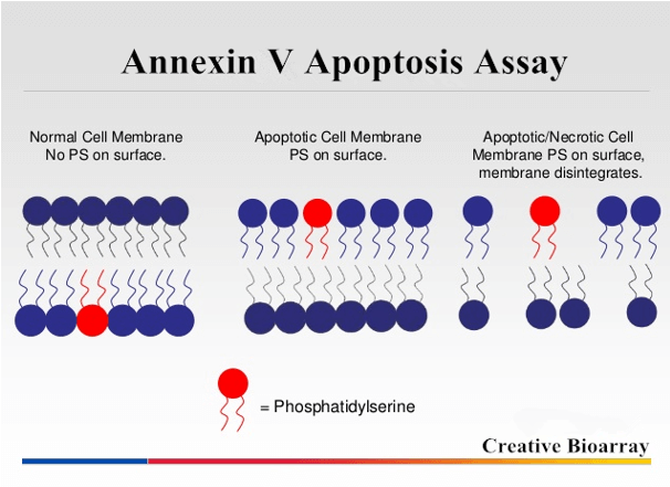 Figure 1. Schematic of Annexin V apoptosis assay.
About the Annexin V-FITC Assay Kits
At Creative Bioarray, our Annexin V-FITC Assay Kit employs FITC-conjugated Annexin V as a probe to detect phosphatidylserine on the outer membrane of apoptotic cells. PI is used as a marker for cell membrane permeability. The kits are useful for assessing mixed populations of apoptotic and necrotic cells by flow cytometry or fluorescence microscopy.
 Figure 2. The expected staining illustration of apoptosis and necrotic cell populations.
Incubation of Cells with Annexin V-FITC and PI
- Inducing cell apoptosis according to your experimental protocol.
- Suspension cells are collected with the culture supernatant, pelleted by centrifugation and washed twice, while adherent cells are harvested by mechanical scraping or gently trypsinizing and washed with serum-containing media.
- Suspension and adherent cells are finally pooled and resuspended in culture medium to a final concentration of 1×10⁶ cells/mL.
- Add Annexin V-FITC and PI to a final concentration of 3 μg/mL and 5 μg/mL, the cells are subsequently incubated for 10 min in the dark.
Flow Cytometry
At least 10,000 cells per sample are analyzed. The wavelength of excitation light is 488 nm, and the emission filters are 515-545 nm (green, FITC) and 600 nm (red, PI). Using electronic compensation to eliminate the bleed through of fluorescence.
Fluorescence Microscopy
Place the cell suspension on a glass slide and cover the cells with a glass coverslip. For adherent cells, they can also be grown directly on a coverslip. The results are observed under a fluorescence microscope by using dual parameter set for FITC and PI. Early apoptosis cells that have bound Annexin V-FITC will show green staining in the plasma membrane. Necrosis cells that have lost membrane integrity will show red staining (PI).
Features
- Easy and fast to operate.
- Wide range for using (both suspension and adherent cells).
- Differentiate early apoptosis from necrosis.
Notes
- All staining procedures must be done in the dark, because Annexin V-FITC and PI are light sensitive.
- It is imperative that samples be analyzed immediately after staining is complete.
- If all samples show strong staining for both Annexin V-FITC and PI, including controls, it is recommended to confirm whether the cells are healthy or incubation time is prolonged.
References
- Devaux P. F. Static and dynamic lipid asymmetry in cell membranes. Biochemistry, 1991, 30: 1163-1173.
- Manon V. E. et al.; Annexin V-affinity assay: a review on an apoptosis detection system based on phosphatidylserine exposure. Cytometry, 1998, 31: 1-9.
- Higgens C. F. Flip-flop: the transmembrane translocation of lipids. Cell, 1994, 79: 393-395.
Cell Services:
Cell Line Testing and Assays:
Explore Other Options