Trypan Blue Staining Assay
One of the earliest and most common methods for measuring cell viability is the trypan blue staining assay. Trypan blue is an azo dye that is cell membrane impermeable and therefore only enters cells with compromised membranes. Upon entry into the cell, trypan blue binds to intracellular proteins thereby rendering the cells a blue color. The trypan blue staining assay allows for a direct identification and enumeration of live (unstained) and dead (blue) cells in a given population.
In this assay, a cell suspension is simply mixed with trypan blue and then visually examined to determine whether cells take up or exclude the dye. In the protocol presented here, a viable cell will have a clear cytoplasm whereas a nonviable cell will have a blue cytoplasm.
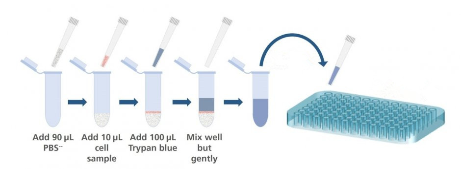 Figure 1. The routine assay process.
Materials
- PBS or serum-free complete medium
- 0.4% trypan blue
Assay Protocol
- Centrifuge the cell suspension being tested for 5 min at 100 × g and discard supernatant.
- Resuspend the cell pellets in 1 mL PBS or serum-free complete medium.
Note: Serum proteins stain with trypan blue and can produce misleading results. Determinations must be made in serum-free solution. - 0.4% trypan blue and cell suspension are mixed. Allow mixture to incubate 3 min at room temperature.
Note: Trypan blue should be sterile filtered before using in order to get rid of particles in the solution that would disturb the counting process.
Mixing can be performed in a well of a microtiter plate or a small plastic tube using 10-20 µL cell suspension and trypan blue. - Apply a drop of the trypan blue/cell mixture to a hemacytometer. Place the hemacytometer on the stage of a binocular microscope and focus on the cells.
Note: Cells should be counted within 3-5 min after mixing with trypan blue, as longer incubation periods will lead to cell death and reduced viability counts. - Count the unstained (viable) and stained (nonviable) cells separately in the hemacytometer. To obtain the total number of viable cells per mL of aliquot, multiply the total number of viable cells by the dilution factor for trypan blue. To obtain the total number of cells per mL of aliquot, add up the total number of viable and nonviable cells.
Note: The aliquot should contain a convenient number of cells to count in a hemacytometer when suspended in 1 mL PBS (e.g., 5 x 10⁵ cells/mL). - Calculate the percentage of viable cells as follows:

Frequently Asked Questions
- Do the cells need to be washed prior to staining with trypan blue solution?
Trypan blue has a greater affinity for serum proteins than for cellular protein. If the background is too dark, cells should be pelleted and resuspended in protein-free medium or salt solution prior to counting.
- Can trypan blue be used to discriminate between apoptotic and necrotic cells?
Trypan blue will stain cells with disrupted membranes, but it cannot differentiate between permeabilized membranes caused by apoptosis or necrosis.
- Trypan blue contains a precipitate. Is it still good to use?
The trypan blue solution is still good to use. It can be warmed to dissolve the precipitate or be filtered to remove the precipitate.
Reference
- Strober W. Trypan blue exclusion test of cell viability. Curr Protoc Immunol, 2001, 21 (1): 1-2.
Cell Services:
Cell Line Testing and Assays:
Explore Other Options