
QualiCell® Human Neuronal Stem Cells (Cortex) -XLC466
- Specification
- Background
- Scientific Data
- Q & A
- Customer Review
Human neuronal stem cells derived from the fetal cerebral cortex—typically isolated at 6–9 weeks of gestation and expanded as neurospheres or adherent cultures—are regionally specified progenitors that preserve the molecular identity of the dorsal telencephalon. Although often referred to as cortical neural stem cells (NSCs), their defining property is the intrinsic commitment to generate cortical projection neurons, interneurons, and glia in proportions that recapitulate human cortical lamination.
Their advantages rest on three pillars. First, stable regional specification – cortical NSCs maintain a PAX6/EMX2‑positive forebrain signature through prolonged passaging. Upon differentiation, they preferentially yield glutamatergic (TBR1+/CTIP2+) pyramidal neurons and calretinin‑positive GABAergic interneurons, a profile distinct from NSCs of spinal or midbrain origin. This regional bias is retained even after xenotransplantation, ensuring predictable graft composition.
Second, exceptional neurogenic capacity – unlike adult or spinal NSCs, cortical NSCs exhibit high proliferative output and generate outer radial glial cells (oRGs), a human‑enriched progenitor population critical for cortical expansion. This enables modelling of human‑specific neurodevelopmental processes—including gyration and layer formation—that are absent in rodent systems.
Third, robust engraftment and functional integration – in stroke and traumatic brain injury models, transplanted cortical NSCs survive, migrate along corticofugal tracts, differentiate into mature neurons that form synaptic contacts with host circuits, and promote behavioural recovery. This therapeutic efficacy, coupled with their amenability to CRISPR‑based gene editing, positions them as an unparalleled platform for disease modelling (e.g., microcephaly, autism, epilepsy) and high‑content drug screening.
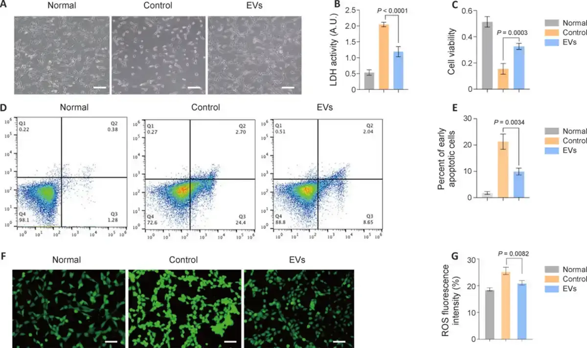
Validation of Human Neural Stem Cell–Derived Extracellular Vesicles Efficacy In Vitro
This study utilized an H2O2-induced oxidative stress model to simulate the pathological environment of ischemic stroke, examining the mechanism of action of human neural stem cell–derived extracellular vesicles (hNSC-EVs) in neuronal repair processes under stressful conditions in vitro. Wang, Jiayi, et al. observed apoptosis-related morphological changes in HT22 cells induced by H2O2 under a microscope, and these morphological changes were partially reversed by treatment with hNSC-EVs (Fig. 1A). Furthermore, lactate dehydrogenase (Fig. 1B) and CCK8 (Fig. 1C) assays showed that hNSC-EVs reduced the damage caused by H2O2-induced oxidative stress, confirming that the hNSC-EVs promoted neuronal repair, reduced toxic damage, and increased cell survival in the context of oxidative stress. Flow cytometry analysis showed a decreased proportion of apoptotic cells in the hNSC-EV group compared with the control group, confirming the role of hNSC-EVs in reversing neuronal apoptosis (Fig. 1D and E). In addition, lower ROS levels were detected in the hNSC-EV group compared with the control group, confirming the antioxidant capacity of hNSC-EVs (Fig. 1F and G).
Ask a Question
Write your own review
- You May Also Need
- Adipose Tissue-Derived Stem Cells
- Human Neurons
- Mouse Probe
- Whole Chromosome Painting Probes
- Hepatic Cells
- Renal Cells
- In Vitro ADME Kits
- Tissue Microarray
- Tissue Blocks
- Tissue Sections
- FFPE Cell Pellet
- Probe
- Centromere Probes
- Telomere Probes
- Satellite Enumeration Probes
- Subtelomere Specific Probes
- Bacterial Probes
- ISH/FISH Probes
- Exosome Isolation Kit
- Human Adult Stem Cells
- Mouse Stem Cells
- iPSCs
- Mouse Embryonic Stem Cells
- iPSC Differentiation Kits
- Mesenchymal Stem Cells
- Immortalized Human Cells
- Immortalized Murine Cells
- Cell Immortalization Kit
- Adipose Cells
- Cardiac Cells
- Dermal Cells
- Epidermal Cells
- Peripheral Blood Mononuclear Cells
- Umbilical Cord Cells
- Monkey Primary Cells
- Mouse Primary Cells
- Breast Tumor Cells
- Colorectal Tumor Cells
- Esophageal Tumor Cells
- Lung Tumor Cells
- Leukemia/Lymphoma/Myeloma Cells
- Ovarian Tumor Cells
- Pancreatic Tumor Cells
- Mouse Tumor Cells