
- Specification
- Background
- Scientific Data
- Q & A
- Customer Review
PG-4 is a feline astrocyte cell line obtained from normal cat brain tissue. The cells show adherent development and morphological features typical with glial cells. PG-4, a continuous feline cell line, has been maintained extensively in vi**tro and used as a host cell culture for investigations of feline-specific diseases and cellular responses to infection.
PG-4 cells are tolerant for various feline viruses and have been utilized to support viral isolation, propagation and infectivity research. They are very susceptible to infection and hence are useful to study the rate of replication of viruses, host cell responses, and the mechanisms influencing virus persistence. In addition to uses in virology, PG-4 cells have been used in studies of cell signaling, cellular metabolism, and interactions between infectious pathogens and neural-derived host cells.
PG-4 cells are of feline origin and thus a species-relevant model for veterinary biomedical research. The cell line can be employed in experimental systems to determine viral pathogenicity, characterize host-pathogen interactions, and examine the biological effects of potential antiviral drugs under controlled laboratory circumstances.
Whole-Genome Analysis Reveals the Probable Donor Cat Phenotype of PG-4 Astrocyte Cells
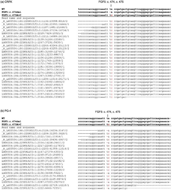
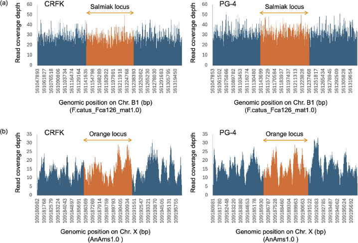
Crandell-Rees Feline Kidney (CRFK) and PG-4 cells are long-established cell lines with undocumented donor origins. Whole-genome sequencing of PG-4 cells identified specific genetic variants linked to coat and iris color. Key findings include a homozygous deletion in the ASIP gene, a compound heterozygous mutation in FGF5 (c.474delT and c.475A>C; Fig. 1), heterozygous mutations in LVRN (c.416C>A and c.2522G>A), and a heterozygous Siamese-color allele in TYR (c.904G>A). No large deletions were found in the KIT-KDR intergenic region or ARHGAP36intron 1 (Fig. 2).
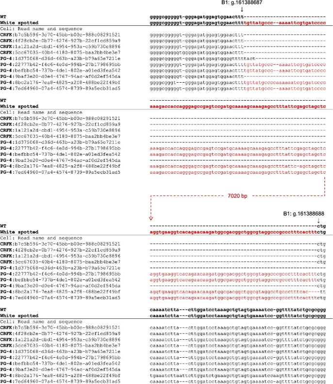
Long-read sequencing revealed that six reads mapped to the KIT-FERV1 locus, with four containing a >7 kbp insertion of the FERV1 homolog (Fig. 3), indicating a heterozygous insertion. While the TYR mutation is recessive and masked by ASIP, the FERV1 insertion is dominant and the FGF5 compound heterozygote causes long fur. Consequently, the PG-4 donor cat likely had white spotting (white and black), unstriped, long fur. The absence of FERV1-LTR or RD114-LTR insertions in PAX3 intron 4 suggests the donor had non-blue irises. These results reconstruct the probable phenotype of the PG-4 donor and highlight the utility of genomic analysis in characterizing historical cell lines.
Ask a Question
Write your own review
Description: CRFK cells were isolated from the cortical portion of the kidneys of a 10-12 week old normal female domestic cat. CRFK are extensively used in feline virus research. Susceptible to herpesvirus and ...
- Adipose Tissue-Derived Stem Cells
- Human Neurons
- Mouse Probe
- Whole Chromosome Painting Probes
- Hepatic Cells
- Renal Cells
- In Vitro ADME Kits
- Tissue Microarray
- Tissue Blocks
- Tissue Sections
- FFPE Cell Pellet
- Probe
- Centromere Probes
- Telomere Probes
- Satellite Enumeration Probes
- Subtelomere Specific Probes
- Bacterial Probes
- ISH/FISH Probes
- Exosome Isolation Kit
- Human Adult Stem Cells
- Mouse Stem Cells
- iPSCs
- Mouse Embryonic Stem Cells
- iPSC Differentiation Kits
- Mesenchymal Stem Cells
- Immortalized Human Cells
- Immortalized Murine Cells
- Cell Immortalization Kit
- Adipose Cells
- Cardiac Cells
- Dermal Cells
- Epidermal Cells
- Peripheral Blood Mononuclear Cells
- Umbilical Cord Cells
- Monkey Primary Cells
- Mouse Primary Cells
- Breast Tumor Cells
- Colorectal Tumor Cells
- Esophageal Tumor Cells
- Lung Tumor Cells
- Leukemia/Lymphoma/Myeloma Cells
- Ovarian Tumor Cells
- Pancreatic Tumor Cells
- Mouse Tumor Cells