Human Mesenchymal Stem Cells-Adult(HMSC-Ad)
- Specification
- Background
- Scientific Data
- Q & A
- Customer Review
Cell Features:
HMSC-Ad are isolated from adult lipoaspirate and are cryopreserved as secondary cells.
HMSC-WJ are isolated from the Wharton's Jelly of human umbilical cord and are cryopreserved as secondary cells.
HMSC-Pre-Adipocyte are isolated from mature adipocytes and are cryopreserved as secondary cells.
HMSC are extensively tested for quality and optimal performance.
Creative Bioarray guarantees performance and quality.
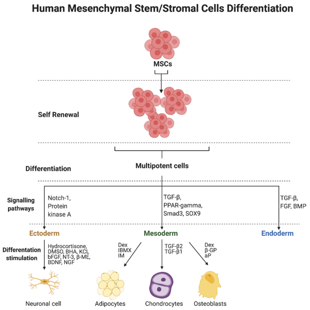
Human Mesenchymal Stem Cells - Adult (HMSC-Ad) are multipotent stem cells derived from adult tissues, most often bone marrow or fat tissue. They are characterized by their ability to self-renew and to differentiate into several lineages, including osteogenic (bone), chondrogenic (cartilage), adipogenic (fat) and other connective tissue lineages.
HMSC-Ad are spindle-shaped, fibroblast-like cells, with a high sensitivity to environmental signals. They secrete a broad range of cytokines and growth factors, which can be involved in tissue repair and regeneration, but also exhibit immune-modulatory functions. This characteristic makes them a unique tool for both regenerative and immunomodulatory research.
HMSC-Ad are used for studies in tissue regeneration, stem cell differentiation, cellular therapies and much more. Their multipotency and ease of cultivation make them a great tool to test new approaches to wound healing, cartilage and bone regeneration, or even for experimental therapies for immune-mediated diseases. In short, HMSC-Ad are the "jack of all trades" of adult stem cells: multipotent, hardy, and very easy to work with in a wide range of experiments.
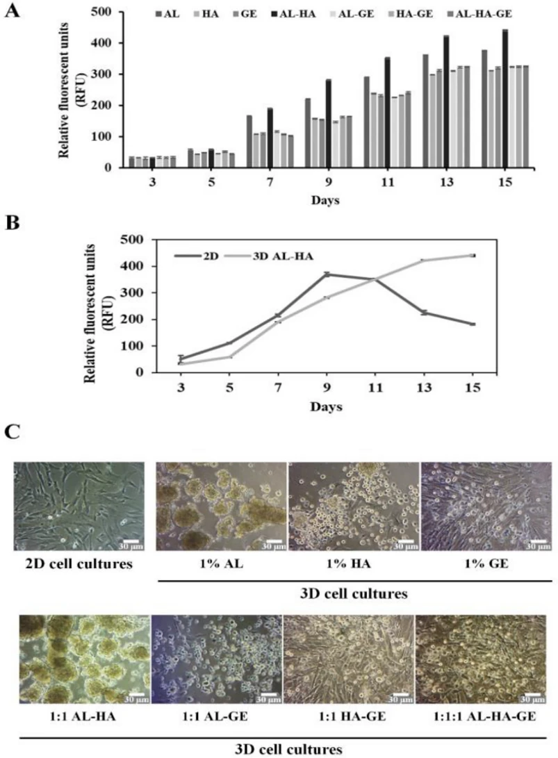
3D Culture of Alginate-Hyaluronic Acid Hydrogel Supports the Stemness of Human Mesenchymal Stem Cells
The three-dimensional (3D) cell culture system is increasingly used to study cell engineering and tissue repair because it closely mimics in vivo microenvironments. In this study, Pangjantuk et al. developed biomaterial hydrogels for 3D native ECM components in hMSCs using photopolymerization. They designed various biomaterial hydrogels (AL, HA, GE, and their combinations) to optimize cell proliferation in 3D hMSC cultures. The proliferation rate of all hydrogels increased with culture time, but after 15 days, AL-HA hydrogels showed the highest proliferation (Fig. 1A). Compared to 2D cultures, 3D AL-HA hydrogels had slower initial growth but higher proliferation after 10 days (Fig. 1B). The population doubling time (PDT) was shorter in 3D AL-HA hydrogels than in 2D cultures. In 2D cultures, hMSCs were spindle-shaped, while in 3D AL-HA hydrogels, they formed cellular spheroids (average size 25-62 µm) after 7 days (Fig. 1C). These results show that 3D AL-HA hydrogels support hMSC proliferation and continuous culture.
Ask a Question
Write your own review
- You May Also Need
- Adipose Tissue-Derived Stem Cells
- Human Neurons
- Mouse Probe
- Whole Chromosome Painting Probes
- Hepatic Cells
- Renal Cells
- In Vitro ADME Kits
- Tissue Microarray
- Tissue Blocks
- Tissue Sections
- FFPE Cell Pellet
- Probe
- Centromere Probes
- Telomere Probes
- Satellite Enumeration Probes
- Subtelomere Specific Probes
- Bacterial Probes
- ISH/FISH Probes
- Exosome Isolation Kit
- Human Adult Stem Cells
- Mouse Stem Cells
- iPSCs
- Mouse Embryonic Stem Cells
- iPSC Differentiation Kits
- Mesenchymal Stem Cells
- Immortalized Human Cells
- Immortalized Murine Cells
- Cell Immortalization Kit
- Adipose Cells
- Cardiac Cells
- Dermal Cells
- Epidermal Cells
- Peripheral Blood Mononuclear Cells
- Umbilical Cord Cells
- Monkey Primary Cells
- Mouse Primary Cells
- Breast Tumor Cells
- Colorectal Tumor Cells
- Esophageal Tumor Cells
- Lung Tumor Cells
- Leukemia/Lymphoma/Myeloma Cells
- Ovarian Tumor Cells
- Pancreatic Tumor Cells
- Mouse Tumor Cells