E.Derm
Cat.No.: CSC-C2920
Species: Equus caballus (Horse)
Source: Skin; Dermis
Morphology: continuous culture, grown as monolayer, morphology fibroblast-like
Culture Properties: monolayer
- Specification
- Background
- Scientific Data
- Q & A
- Customer Review
Tissue: dermis
E.Derm is an equine dermal fibroblast cell line obtained from the skin of a healthy adult American Quarter Horse. The cell line is well known as NBL-6 and has been extensively employed in veterinary and comparative biomedical research. As a dermis-derived fibroblast model, E.Derm cells exhibit a typical spindle-shaped morphology and grow as an adherent monolayer under standard culture conditions. The cells retain several properties of connective tissue fibroblasts including formation of extracellular matrix, participation in tissue remodeling and reactivity to environmental cues.
E.Derm has been widely used in studies of infectious illnesses of equine origin, mainly as a host cell model in studies of viral transmission and host-pathogen interaction. It is permissive for infection with a number of horse viruses including equine influenza virus, equine herpesviruses and equine toroviruses. The cell line is a helpful tool to study viral replication, cellular responses to infection and mechanisms of viral adaptability. In addition to uses in virology, E.Derm cells have been used in studies of innate immune signaling, cytokine responses, and comparative host susceptibility to infectious pathogens.
EIV/63 and EIV/2003 Exhibit Significant Differences in Replication Kinetics, Cell-To-Cell Spread and Susceptibility to Interferon In Vitro
To study the effects of viral evolution on fitness, the researchers performed controlled infections with two H3N8 EIV strains isolated 40 years apart. Mamalian cell lines and horse tracheal explants were infected and viral replication, interferon resistance, cell-to-cell dissemination and tissue damage were assayed. Transcriptomics were used to profile host responses, while image analysis was used to quantify epithelial lesions.
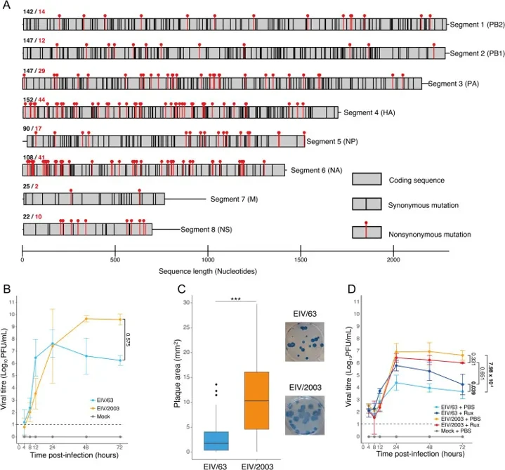
The replication dynamics of EIV/1963 and EIV/2003 were studied in mammalian cells to determine adaptive changes between evolutionarily divergent equine influenza viruses (EIV). Infections were performed in both Madin-Darby Canine Kidney (MDCK) cells and interferon-competent equine dermal fibroblasts (E.Derm), the latter being a more physiologically relevant model. Both viruses replicated with similar kinetics in MDCK cells, however, EIV/2003 reached peak titres three logs greater than EIV/1963 at 48 hpi (Fig. 1B). EIV/2003 also developed considerably larger plaques indicating increased cell to cell dissemination (Fig. 1C, p< 10-17). In E.Derm cells, EIV/2003 always beat EIV/1963 and achieved titers three logs higher from 24 hpi (Fig. 1D, p= 7.98 × 10⁻⁴). EIV/1963 replication was considerably increased following treatment with the JAK1/2 inhibitor, ruxolitinib (p= 0.039) and was comparable to that of EIV/2003. By contrast, ruxolitinib did not impact EIV/2003 replication (p= 0.331). These findings show that the present EIV/2003 lineage is more fit and more resistant to the horse type I interferon response relative to the historical EIV/1963 strain.
Ask a Question
Write your own review
- You May Also Need
- Adipose Tissue-Derived Stem Cells
- Human Neurons
- Mouse Probe
- Whole Chromosome Painting Probes
- Hepatic Cells
- Renal Cells
- In Vitro ADME Kits
- Tissue Microarray
- Tissue Blocks
- Tissue Sections
- FFPE Cell Pellet
- Probe
- Centromere Probes
- Telomere Probes
- Satellite Enumeration Probes
- Subtelomere Specific Probes
- Bacterial Probes
- ISH/FISH Probes
- Exosome Isolation Kit
- Human Adult Stem Cells
- Mouse Stem Cells
- iPSCs
- Mouse Embryonic Stem Cells
- iPSC Differentiation Kits
- Mesenchymal Stem Cells
- Immortalized Human Cells
- Immortalized Murine Cells
- Cell Immortalization Kit
- Adipose Cells
- Cardiac Cells
- Dermal Cells
- Epidermal Cells
- Peripheral Blood Mononuclear Cells
- Umbilical Cord Cells
- Monkey Primary Cells
- Mouse Primary Cells
- Breast Tumor Cells
- Colorectal Tumor Cells
- Esophageal Tumor Cells
- Lung Tumor Cells
- Leukemia/Lymphoma/Myeloma Cells
- Ovarian Tumor Cells
- Pancreatic Tumor Cells
- Mouse Tumor Cells