BALB/c Mouse Bone Marrow Mesenchymal Stem Cells
- Specification
- Background
- Scientific Data
- Q & A
- Customer Review
Mouse Bone Marrow Mesenchymal Stem Cells are negative for bacteria, yeast, fungi, and mycoplasma. Cells can be expanded on a multiwell culture plate ready for experiments under the cell culture conditions specified by Creative Bioarray. Repeated freezing and thawing of cells is not recommended.
Each batch of Mouse Bone Marrow Mesenchymal Stem Cells are tested for expression of markers using antibodies, CD44, Sca-1 and CD29 by flow cytometry.
Standard biochemical procedures performed with cell cultures include RT-PCR, Western blotting, immunoprecipitation, immunofluorescent staining, flow cytometry or generating cell derivatives for desired research applications.
BALB/c Mouse Bone Marrow Mesenchymal Stem Cells (BM-MSCs) are harvested directly from the bone marrow of BALB/c mice. These primary multipotent stromal cells are commonly employed in stem cell research, immunology, and regenerative medicine applications. BALB/c Mouse BM-MSCs maintain key features of traditional MSCs such as plastic adherence, fibroblast-like morphology, and the ability to proliferate extensively in standard culture conditions. BM-MSCs can differentiate into osteoblasts, adipocytes, chondrocytes, and other cell types upon cultivation in appropriate lineage-specific differentiation media. In addition, they maintain the typical MSC phenotype by expressing cell surface markers such as CD29, CD44, and Sca-1 and lack hematopoietic markers such as CD34 and CD45. BALB/c Mouse Bone Marrow Mesenchymal Stem Cells provide a consistent and reproducible source of MSCs for use in your research.
Derived from an inbred strain, these cells ensure superior genetic uniformity for use in syngeneic transplant applications and immunomodulation studies. Researchers commonly utilize these cells to study tissue regeneration, inflammation, tumor-stroma interactions, and cell therapy development. BALB/c Mouse BM-MSCs exhibit stable performance, high viability, and wide applicability in preclinical studies.
Effect of High Mobility Group Box-1 on Mitochondrial Transfer
Mitochondria are crucial for cell metabolism and tissue survival, and their transfer between cells can impact recipient cell fate. BM-MSCs have been reported to transfer mitochondria to cancer cells, rescuing mitochondrial dysfunction, but the mechanisms are unclear. Sasaki et al. investigated the humoral factors and mechanisms involved in mitochondrial transfer (MT) from BM-MSCs to colorectal cancer (CRC) cells.
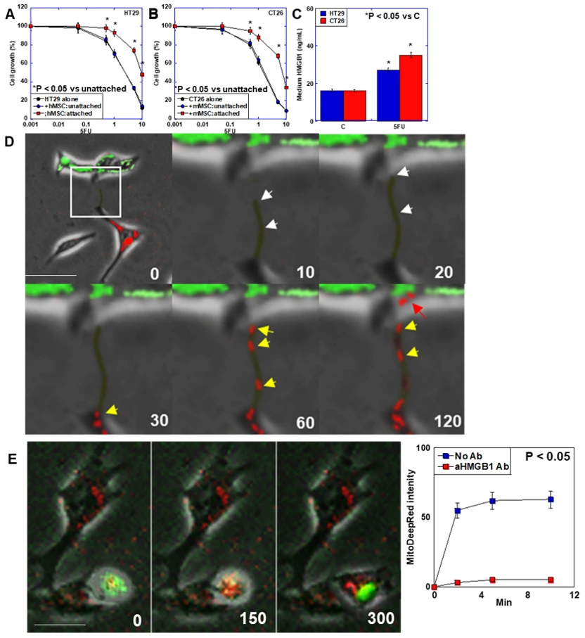
To assess the relationship between colorectal cancer (CRC) cells and bone marrow mesenchymal stem cells (BM-MSCs), co-culture experiments were conducted under both unattached and attached conditions (Fig. 1A, B). In the unattached condition, HT29 and CT26 CRC cells showed cell growth at the same levels to those in non-cocultured cells, whereas both CRC cells showed enhanced cell growth in the attached condition. CRC cells treated with 5-FU increased the secretion of HMGB1 into the cultured medium (Fig. 1C). To confirm MT from BM-MSCs to CRC cells, CRC cells (PKH67 labeled) and BM-MSCs (mitochondria labeled with Mito Deep red) were cocultured (Fig. 1D, E). Under 5FU treatment, Mouse BM-MSC (mBM-MSC) elongated TNT to the CT26 CRC cell. Mitochondria of mBM-MSCs were transferred to the CT26 cell through the TNT (Fig. 1D). The mMSC TNT reached the CT26 cell at 20 s. The mMSC mitochondria entered into the TNT at 30 s. The mMSC mitochondria reached the CT26 cell cytoplasm. The hBM-MSCs attached to the HT29 CRC cell. Mitochondria of hBM-MSCs were transferred to the HT29 cell temporally (Fig. 1E).
Ask a Question
Write your own review
- You May Also Need
- Adipose Tissue-Derived Stem Cells
- Human Neurons
- Mouse Probe
- Whole Chromosome Painting Probes
- Hepatic Cells
- Renal Cells
- In Vitro ADME Kits
- Tissue Microarray
- Tissue Blocks
- Tissue Sections
- FFPE Cell Pellet
- Probe
- Centromere Probes
- Telomere Probes
- Satellite Enumeration Probes
- Subtelomere Specific Probes
- Bacterial Probes
- ISH/FISH Probes
- Exosome Isolation Kit
- Human Adult Stem Cells
- Mouse Stem Cells
- iPSCs
- Mouse Embryonic Stem Cells
- iPSC Differentiation Kits
- Mesenchymal Stem Cells
- Immortalized Human Cells
- Immortalized Murine Cells
- Cell Immortalization Kit
- Adipose Cells
- Cardiac Cells
- Dermal Cells
- Epidermal Cells
- Peripheral Blood Mononuclear Cells
- Umbilical Cord Cells
- Monkey Primary Cells
- Mouse Primary Cells
- Breast Tumor Cells
- Colorectal Tumor Cells
- Esophageal Tumor Cells
- Lung Tumor Cells
- Leukemia/Lymphoma/Myeloma Cells
- Ovarian Tumor Cells
- Pancreatic Tumor Cells
- Mouse Tumor Cells