Rat Renal Glomerular Endothelial Cells
Cat.No.: CSC-C9377W
Species: Rat
Source: Kidney
Morphology: Polygonal
Cell Type: Endothelial Cell
- Specification
- Background
- Scientific Data
- Q & A
- Customer Review
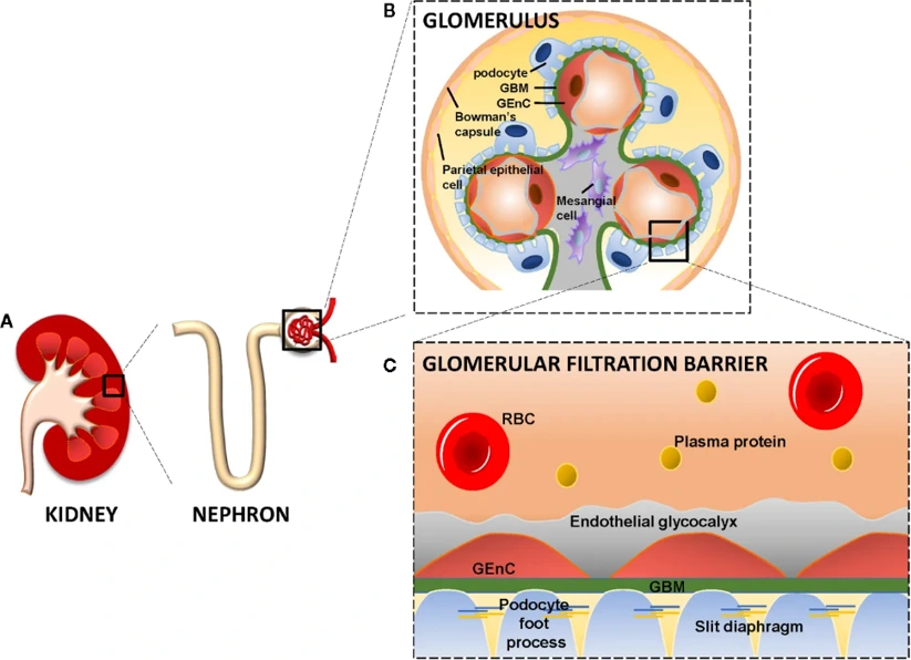
Rat Renal Glomerular Endothelial Cells (RGECs) are endothelial cells isolated from rat renal glomerular capillaries. RGECs line glomerular capillaries and along with the glomerular basement membrane and podocytes make up the glomerular filtration barrier. These cells selectively filter plasma across the glomerular capillaries. Glomerular endothelial cells function as an endothelial barrier that prevents plasma proteins and blood cells from leaking out of capillaries but allows free movement of water and small solutes during normal kidney filtration through fenestrations.
RGECs typically display a cobblestone morphology observed of most endothelial cells when maintained in culture. Additionally, these cells express endothelial cell specific markers including CD31 (PECAM-1), von Willebrand factor (vWF) and vascular endothelial cadherin (VE-cadherin). They are also exhibit endothelial cell functions such as Ac-LDL uptake, nitric oxide production, and ability to form capillary-like structures in vitro. In vitro studies commonly use RGECs to study kidney microvascular cell biology as well as aspects of glomerular endothelial barrier function. RGECs have been used to study endothelial dysfunction, inflammatory signaling pathways, oxidative stress, angiogenesis, and nephrotoxicity.
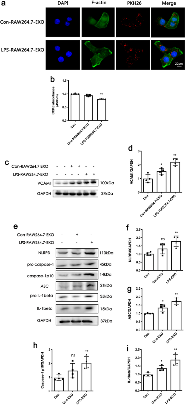
EXOs from LPS-Stimulated Macrophages Can Induce the Damage of Glomerular Endothelial Cell In Vitro
Sepsis-associated AKI is associated with sepsis related mortality. Activation of macrophages and endothelial injury play a role in AKI, however the mechanism remains poorly defined. LPS stimulated macrophage derived exosomes were co-incubated with rat glomerular endothelial cells (RGECs) and markers of injury were assessed in vitro. The ASM inhibitor amitriptyline was used to identify ASM involvement. Confocal microscopy confirmed uptake of RAW264.7 derived exosomes by RGECs (Fig.1a). Co-incubation of RGECs with LPS stimulated macrophage derived exosomes decreased cell viability and induced VCAM-1 expression in RGECs (Fig. 1b, d). LPS stimulated macrophage derived exosomes also activated NLRP3 inflammasomes in RGECs, determined by upregulation of NLRP3, ASC, Caspase-1 and IL-1β (Fig. 1e-h).
Ask a Question
Write your own review

Description: The thoracic aorta is located in the chest cavity and gives off arteries that branch to the esophagus, pericardium, lungs, and trachea. The thoracic aorta can be subdivided into the ascending aorta, ...

Description: Rat Podocytes are isolated from normal rat kidney. The cells are characterized by immunofluorescence with antibodies specific to podocin, Ang1, Nephrin, ACTN4, NPHS2. T25 flasks is required for cell ...

Description: Rat Bronchial Smooth Muscle Cells are isolated from normal rat bronchi tissue. Rat Bronchial Smooth Muscle Cells are characterized by immunofluorescence with antibodies specific to alpha-actin. T25 ...

Description: Guinea Pig Endothelial Cells from Creative Bioarray are isolated from guinea pig tissue. Prior to shipping, cells at passage 2 are detached from flasks and immediately cryopreserved in vials. Each ...

Description: Rat Lung Epithelial Cells are isolated from normal rat lung tissue. The cells are characterized by immunofluorescence with antibodies specific to CK-18, CK-19. T25 flasks is required for cell ...

Description: Rat Vein Endothelial Cells from Creative Bioarray are isolated from inferior vena cava tissue of 6-8 week old laboratory Sprague-Dawley rat. Rat Vein Endothelial Cells are grown in T75 tissue culture ...