Rat Microglia
Cat.No.: CSC-C1800
Species: Rat
Source: Brain
Cell Type: Microglia; Glial Cell
- Specification
- Background
- Scientific Data
- Q & A
- Customer Review
Microglia are the primary immune cells of the CNS of the rat. They are developmentally derived from mesodermic progenitors and become the predominant mediators of innate immunity in the brain and spinal cord early in life. Rat Microglia play an important role in CNS homeostasis.
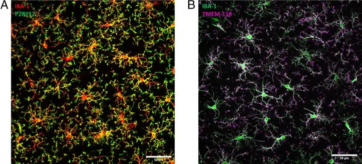
Microglia are ramified in the resting state and express typical microglial markers such as Iba1, CD11b, CD68 and CX3CR1. They can become activated in the presence of inflammation, pathogens or neural damage. These activated microglia can release cytokines, chemokines, reactive oxygen species, as well as neurotrophic factors. These cells in culture are widely used to study neuroimmune interactions, neuroinflammation, and CNS pathologies. They are utilized in studying neurodegenerative diseases, brain trauma, stroke, infection and neurotoxicity. These cells can also be used to study inflammatory signaling pathways as well as microglia-neuron signaling. Rat models are often used to study neurological diseases; therefore, rat microglia provide an easily accessible in vivo system to study CNS immune regulation and test therapeutic interventions.
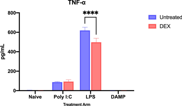
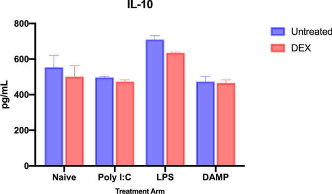
Dexmedetomidine Alters the Inflammatory Cytokine Profile of Rat Microglia In Vitro
Microglia are responsible for neuroinflammation after neurologic insults such as traumatic brain injury (TBI). They undergo significant changes in cytokine expression, metabolism, and immunophenotype after injury. Dexmedetomidine (DEX), an α2 adrenergic agonist frequently used to sedate critically ill patients with TBI, may attenuate inflammatory microglia phenotypes. Primary rat microglia were activated with LPS, Poly I: C, or TBI-derived damage-associated molecular patterns (DAMP) then treated with Dex. Supernatants and cells were collected after 24 h for analysis.
This study found LPS and Poly I: C to significantly increase TNFα (p < 0.0001 and p = 0.0005, respectively), while DAMP had no significant effect. Treatment with DEX reduced TNFα released by LPS-stimulated microglia (p < 0.0001; Fig. 1) but did not affect TNFα in naïve, Poly I:C or DAMP stimulated microglia. DEX significantly decreased TNFα across all cultures (p < 0.0001). LPS caused a significant increase in IL-10 (p = 0.0002) while Poly I:C and DAMP caused a non-significant decrease. Treatment with DEX reduced IL-10 production by LPS (p = 0.0039) and Poly I: C (p = 0.0221) stimulated microglia, but not naïve or DAMP-stimulated microglia (Fig. 2). DEX significantly decreased IL-10 expression across all cultures (p < 0.0001).
Rat Microglia are primary cells, not a cell line.
Ask a Question
Write your own review
- You May Also Need

Description: The thoracic aorta is located in the chest cavity and gives off arteries that branch to the esophagus, pericardium, lungs, and trachea. The thoracic aorta can be subdivided into the ascending aorta, ...

Description: Rat Podocytes are isolated from normal rat kidney. The cells are characterized by immunofluorescence with antibodies specific to podocin, Ang1, Nephrin, ACTN4, NPHS2. T25 flasks is required for cell ...

Description: Rat Bronchial Smooth Muscle Cells are isolated from normal rat bronchi tissue. Rat Bronchial Smooth Muscle Cells are characterized by immunofluorescence with antibodies specific to alpha-actin. T25 ...

Description: Guinea Pig Endothelial Cells from Creative Bioarray are isolated from guinea pig tissue. Prior to shipping, cells at passage 2 are detached from flasks and immediately cryopreserved in vials. Each ...

Description: Rat Lung Epithelial Cells are isolated from normal rat lung tissue. The cells are characterized by immunofluorescence with antibodies specific to CK-18, CK-19. T25 flasks is required for cell ...

Description: Rat Vein Endothelial Cells from Creative Bioarray are isolated from inferior vena cava tissue of 6-8 week old laboratory Sprague-Dawley rat. Rat Vein Endothelial Cells are grown in T75 tissue culture ...