
Rat Astrocytes
Cat.No.: CSC-C1797
Species: Rat
Source: Brain
Cell Type: Astrocyte; Glial Cell
- Specification
- Background
- Scientific Data
- Q & A
- Customer Review
RA are isolated from day 2 rat cerebral cortex. RA are cryopreserved at secondary culture and delivered frozen. Each vial contains >1 x 10^6 cells in 1 ml volume. RA are characterized by immunofluorescent method with antibody to GFAP. RA are negative for mycoplasma, bacteria, yeast and fungi. RA are guaranteed to further expand for 5 population doublings in the condition provided by Creative Bioarray.
Rat astrocytes are primary astrocyte cell cultures that have been isolated from rat brain tissue (usually cortex or hippocampus) during infancy. Typically, the resulting culture is predominately Type I astrocytes, which have a flat, polygonal, "fried-egg" appearance. They express definitive marker proteins such as glial fibrillary acidic protein (GFAP) and S100β. These cells readily proliferate in culture and will usually grow to confluence, allowing for easy generation of astrocyte/neuron co-cultures.
Rat astrocytes are used extensively in neurobiology because rats are the predominant model organism used, making this cell type both standard and physiologically relevant. These cells are used to understand basic physiology and disease pathology. Major uses include: blood brain barrier modeling (in endothelial cell co-culture), neuroinflammation (reactivity, cytokine secretion), neuronal support and synapse modulation, excitotoxicity, and many neurodegenerative diseases (Alzheimer's, Parkinson's, stroke). As with any primary cell, there is donor variability and passage-dependent changes to phenotype and cellular behavior, as well as a limited lifespan. However, they are more physiologically relevant than many immortalized astrocyte cell lines.
ATP Contents of Cultured Astrocytes after 24 h Incubation with or without Glucose
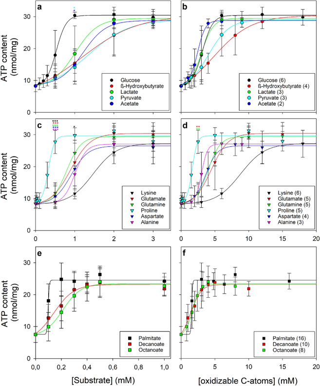
Brain astrocytes maintain high ATP levels for hours after glucose deprivation by mobilizing endogenous substrates, but cellular ATP drops to ~30% of initial content within 24 h. Harders et al. investigated their metabolic flexibility by screening exogenous substrates for ATP maintenance, identifying fatty acids, monocarboxylates, purine nucleosides, and specific amino acids-particularly proline-as potent alternative fuels supporting mitochondrial ATP regeneration.
To confirm previous findings and establish baseline conditions, they analyzed 39 experiments from 30 independent cultures. Initial specific ATP content (30 ± 5 nmol/mg) decreased by 72% to 8 ± 2 nmol/mg after 24 h glucose deprivation, whereas 3 mM glucose maintained 87 ± 13% of initial ATP. Neither condition caused cell toxicity, as shown by low extracellular LDH activities. Glucose concentration-dependent analysis revealed half-maximal and maximal ATP maintenance at ~0.5 mM and 1 mM (Fig. 1a), respectively.
Ask a Question
Write your own review

Description: The thoracic aorta is located in the chest cavity and gives off arteries that branch to the esophagus, pericardium, lungs, and trachea. The thoracic aorta can be subdivided into the ascending aorta, ...

Description: Rat Podocytes are isolated from normal rat kidney. The cells are characterized by immunofluorescence with antibodies specific to podocin, Ang1, Nephrin, ACTN4, NPHS2. T25 flasks is required for cell ...

Description: Rat Bronchial Smooth Muscle Cells are isolated from normal rat bronchi tissue. Rat Bronchial Smooth Muscle Cells are characterized by immunofluorescence with antibodies specific to alpha-actin. T25 ...

Description: Guinea Pig Endothelial Cells from Creative Bioarray are isolated from guinea pig tissue. Prior to shipping, cells at passage 2 are detached from flasks and immediately cryopreserved in vials. Each ...

Description: Rat Lung Epithelial Cells are isolated from normal rat lung tissue. The cells are characterized by immunofluorescence with antibodies specific to CK-18, CK-19. T25 flasks is required for cell ...

Description: Rat Vein Endothelial Cells from Creative Bioarray are isolated from inferior vena cava tissue of 6-8 week old laboratory Sprague-Dawley rat. Rat Vein Endothelial Cells are grown in T75 tissue culture ...