Rabbit Cardiomyocytes
Cat.No.: CSC-C5257S
Species: Rabbit
Source: Heart
Cell Type: Cardiomyocyte
- Specification
- Background
- Scientific Data
- Q & A
- Customer Review
Rabbit cardiomyocytes from Creative Bioarray are isolated from rabbit heart tissue. The method we use to isolate rabbit cardiomyocytes was developed based on a combination of established and our proprietary methods. The rabbit cardiomyocytes from Creative Bioarray are characterized by immunofluorescence with antibodies specific to myosin heavy chain. Each vial contains 0.5x10^6 cells per ml and is delivered frozen.
"Rabbit Cardiomyocytes" primarily refer to isolated primary cells obtained from the ventricular or atrial tissue of rabbit hearts, most commonly from neonatal, juvenile, or adult New Zealand White rabbits. Rabbit cardiomyocytes are terminally differentiated and thus will not replicate under culture conditions. They maintain their contractile phenotype and rod-shaped morphology for a short time in vitro (usually up to 7-10 days).
Cells are usually isolated by enzymatic (collagenase) digestion of cardiac tissue. Although culture consists of mixed-cell populations, steps can be taken to enrich for cardiomyocytes. They display characteristic cardiac properties such as spontaneous or rhythmic contraction, negative force-frequency relationship, and cardiac-specific protein expression (troponin I, α-actinin, connexin 43). Furthermore, rabbit cardiomyocytes display an action potential profile that is more representative of the human heart compared to rodent models. For this reason, they are widely used for electrophysiology/pharmacology purposes. Rabbit cardiomyocytes have served as the gold-standard model for cardiac safety pharmacology (i.e. investigation of drug-induced cardiac arrhythmias such as Torsades de Pointes), investigation into the pathophysiology of heart failure and ischemia/reperfusion, as well as studies into contractile function. Rabbit cardiomyocytes provide a balance between lower animal models such as cellular lines and higher models such as whole-heart and in vivomodels.
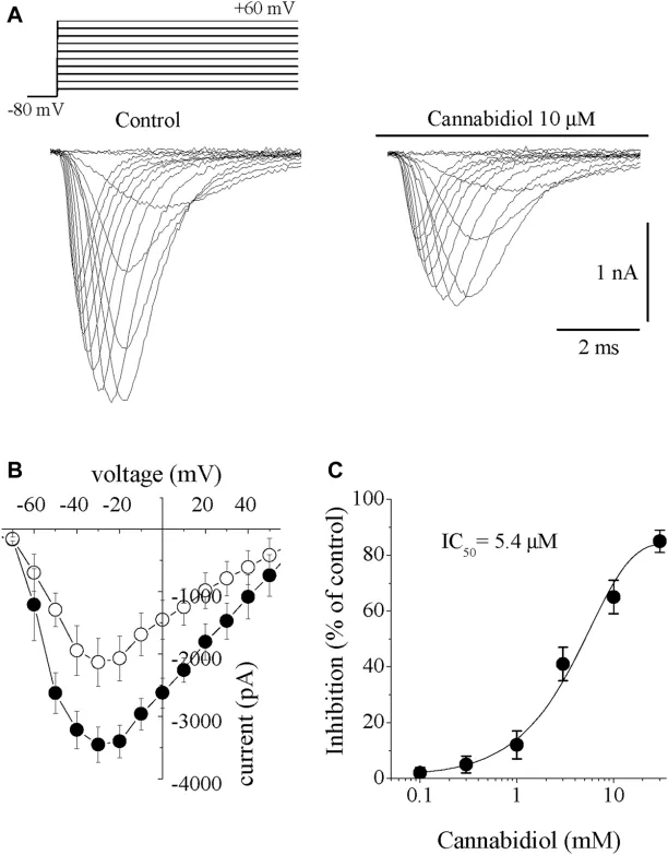
The Effects of Cannabidiol on the Na Channels (INa+)
Cannabidiol (CBD), the prevalent non-psychotropic cannabinoid produces anti-nociceptive, anti-psychotic, anti-convulsant and cardiovascular actions. Here, Isaev et al. examined the effects of CBD on major ion currents in rabbit ventricular myocytes.
Inward sodium current (INa) was elicited by 10 mV step depolarizations from -80 mV holding potential. INa activated at -50 mV, peaked at -30 mV, and reversed at ~+60 mV. CBD (0.3-30 µM) caused concentration-dependent INa suppression detectable at 2-3 min, reaching steady-state at 10-15 min with partial recovery (n = 6). Traces before and after 10 µM CBD are shown (Figure 1A). CBD inhibited INa without altering the I-V relationship, threshold, peak, or reversal potentials (Fig. 1B). The concentration-response curve yielded an IC50 of 5.4 µM and Hill coefficient of 2.6 (n = 6-8; Fig. 1C).
Ask a Question
Write your own review
Description: Rabbit Hepatocytes are derived from the liver of New Zealand White Rabbit.
Description: The aortic arch is the top part of the main artery carrying blood away from the heart. It is the connection between the ascending and descending aorta, and its central part is formed by the left 4th ...
Description: The synovium secretes synovial fluid, which plays an important role in joint movement. The normal synovium has two layers, a thin cellular layer (luminal layer) and a vascular layer (subintima). ...
Description: The pituitary gland is an important endocrine gland in the body that secretes growth hormone and adrenocorticotropic hormone. It plays an important role in the growth and development of the body, ...

Description: The oral epthelial cells are responsible for important functions, like the primary protection of oral mucosa against external aggressions building a mechanical barrier against microorganisms, ...
Description: The carotid arteries are major blood vessels in the neck that supply blood to the brain, neck, and face. There are two carotid arteries, one on the right and one on the left. In the neck, each ...