C57BL/6 Mouse Primary Lung Fibroblasts
Cat.No.: CSC-C1880
Species: Mouse
Source: Lung
Morphology: Multipolar
Cell Type: Fibroblast
- Specification
- Background
- Scientific Data
- Q & A
- Customer Review
C57BL/6 mouse primary lung fibroblasts are isolated from the lung parenchyma of the widely adopted C57BL/6 inbred strain, representing a gold-standard model for pulmonary research. These cells are essential mediators of extracellular matrix (ECM) turnover, secreting collagen, fibronectin, and matrix metalloproteinases, and play pivotal roles in wound healing, inflammation, and fibrotic remodeling. Unlike immortalized cell lines, primary lung fibroblasts retain physiological responsiveness, native signaling pathways, and donor-specific genetic background, ensuring superior predictive power for in vivo translational outcomes.
The C57BL/6 genetic background confers distinct commercial advantages: fully sequenced genome, low inter-animal variability, consistent immune and fibrotic responses, and extensive validation in respiratory disease models, including idiopathic pulmonary fibrosis (IPF), chronic obstructive pulmonary disease (COPD), asthma, and acute lung injury. Primary cells from this strain faithfully recapitulate pathological features such as TGF-β1-induced myofibroblast differentiation, excessive ECM deposition, and aberrant proliferation.
Offered as cryopreserved, high-viability (>90%) preparations with defined phenotype (vimentin+, collagen I+, α-SMA inducible), these cells provide a reproducible, scalable platform for high-content drug screening, anti-fibrotic candidate testing, mechanobiology studies, and toxicity assessment. Their batch-to-batch consistency reduces animal usage and accelerates preclinical discovery, making them an enabling tool for pharmaceutical development, regenerative medicine, and respiratory disease modeling in commercial settings.
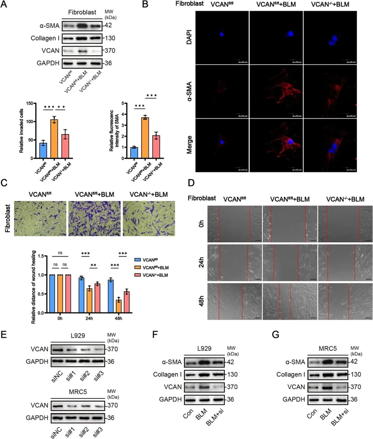
Specific Knockout of VCAN in Fibroblasts Attenuates Lung Fibrosis
This study aimed to elucidate the role and mechanism of versican (VCAN) in promoting pulmonary fibrosis (PF) and to identify therapeutic targets for PF. Given that fibroblasts are the main effector cells in the process of lung fibrosis, we investigated whether changes in VCAN expression during lung fibrosis were associated with fibroblast activation.
Primary lung fibroblasts were isolated from wild-type C57BL/6 mice along with VCANfl/fl (control) and VCAN-/- (fibroblast-specific knockout) mice, and their activation levels were examined. The results of the WB experiments showed that bleomycin (BLM) significantly induced fibroblasts to express α-SMA and Collagen. However, in mice with specific VCAN knockout in fibroblasts, the ability of BLM to induce α-SMA and Collagen expression was significantly decreased (Fig. 1A). Immunofluorescence experiments for α-SMA also showed that BLM-induced α-SMA expression in fibroblasts was significantly lower in mice with specific knockout of VCAN than in mice treated with BLM alone (Fig. 1B). Activated fibroblasts showed significant migration capacity. Transwell and scratch assays demonstrated that BLM significantly increased the migratory capacity of fibroblasts. However, the fibroblast migration ability significantly decreased in the VCAN knockout group compared to that in the BLM group (Fig. 1C-D).
To further validate our experimental results, we used human and mouse-derived fibroblast cell lines to verify the effect of VCAN on fibroblast activation, the knockdown efficiency was validated, and those with high efficiency were selected for subsequent experiments (Fig. 1E). WB results showed that VCAN knockdown significantly inhibited BLM-induced fibroblast expression of α-SMA and Collagen (Fig. 1F, G). These results indicated that VCAN plays an important role in the activation of lung fibroblasts.
Ask a Question
Write your own review


Description: C57BL/6-GFP Mouse Skeletal Muscle Microvascular Endothelial Cells from Creative Bioarray are isolated from C57BL/6-Tg (CAG-EGFP) 1Osb/J mouse skeletal muscle tissue of pathogen-free laboratory mice. ...

Description: eNOS KO Mouse Stomach Epithelial Cells from Creative Bioarray are isolated from stomach tissue of pathogen-free laboratory mice. eNOS KO Mouse Stomach Epithelial Cells are grown in a T25 tissue ...
Description: eNOS KO Mouse Liver Fibroblasts from Creative Bioarray are isolated from liver tissue of pathogen-free laboratory mice. eNOS KO Mouse Liver Fibroblasts are grown in T75 tissue culture flasks ...
Description: C57BL/6-GFP Mouse Corneal Epithelial Cells from Creative Bioarray are isolated from C57BL/6-GFP-Tg(CAG-EGFP)1Osb/J mouse corneal tissue of pathogen-free laboratory mice. C57BL/6-GFP Mouse Corneal ...

Description: BALB/c Mouse Retinal Microvascular Endothelial Cells from Creative Bioarray are isolated from retinal tissue of pathogen-free laboratory mice. BALB/c Mouse Retinal Microvascular Endothelial Cells are ...