
Immortalized Human Renal Proximal Tubule Cells-hTERT
Cat.No.: CSC-I0721Z
Species: homo sapiens
Source: Kidney
Cell Type: Epithelial Cells
- Specification
- Background
- Scientific Data
- Q & A
- Customer Review
free from contaminations (bacteria incl. mycoplasma, fungi, HIV, HAV, HBV, HCV, Parvo-B19) and cross-contaminations
Immortalized Human Renal Proximal Tubule Cells-hTERT (RPTEC-hTERT) are human renal proximal tubular epithelial cells immortalized by stable expression of human telomerase reverse transcriptase (hTERT). This method of immortalization allows cells to bypass senescence without introduction of viral oncogenes (SV40, for example).
RPTEC-hTERT cells are morphologically similar to other proximal tubule epithelial cells demonstrating an epithelial cobblestone appearance when grown as a polarized monolayer with tight junctions. RPTEC-hTERT express cell surface markers typical of proximal tubule cells such as E-cadherin, cytokeratin 18, aquaporin-1, megalin (LRP2), Na⁺/K⁺-ATPase as well as drug transporters OAT1, OAT3, OCT2, and P-glycoprotein which allows use of these cells in renal transport and nephrotoxicity studies. These cells have been shown to actively uptake/transport organic cations and organic anions and secrete these compounds. Exposure of RPTEC-hTERT cells to nephrotoxicants also induces physiologically relevant responses. Applications of these cells include renal drug transport studies, xenobiotic metabolism, nephrotoxicity screenings, oxidative stress, fibrosis signaling pathways, and kidney disease models.
Guanine Quadruplex Mediated Pausing Regulates Mitochondrial Function
DNA G4 structures are established regulators of transcription yet roles for RNA G4s in regulating mitochondrial transcription have not been investigated. Snyder et al. sought to determine if RNA G4s regulate mitochondrial RNA polymerase (POLRMT) pausing and mitochondrial function.
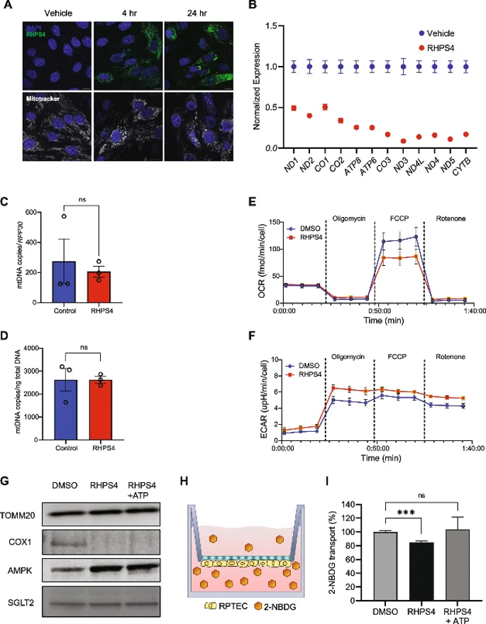
They tested the functional consequences of G4 mediated POLRMT pausing in immortalized human renal proximal tubule cells (RPTEC) due to the kidney's metabolic requirements. Treatment with RHPS4 for 24h resulted in accumulation of POLRMT in mitochondria in a time dependent manner (Fig. 1A) and significantly reduced expression of all 12 heavy strand-encoded polycistronic mRNAs, including MT-CO1 (Fig. 1B). This occurred while maintaining mtDNA content (Fig. 1C, D). Expression level of each gene was negatively correlated with distance from the transcription start site, further supporting the idea that POLRMT accumulation at G4 sites inhibits distal transcription. Mitochondria-encoded proteins are required for respiration so they measured oxygen consumption. RHPS4 significantly inhibited O₂ consumption (Fig. 1E) and increased compensatory glycolysis (Fig. 1F). Thus, stabilizing G4s to induce POLRMT pausing impaired mitochondrial gene expression and cellular energy production. RHPS4 decreased mitochondria-encoded COX1 protein without changing nuclear-encoded TOMM20 or mitochondria abundance in RPTECs grown on transwell inserts (Fig. 1G). This caused an imbalance in cellular energy that activated AMPK (Fig. 1G).
Ask a Question
Write your own review

Description: Nasal epithelial cells form the outermost protective layer against environmental factors. They clean, humidify, and warm inhaled air. They produces mucus, which binds particles that are subsequently ...
Description: Immortalized Human Corneal Epithelial Cells-SV40 have been obtained immortalizing Human Corneal Epithelial Cells with Lenti-SV40 Lentivirus. Immortalized cells were controlled passaging side by side ...
Description: Immortalized Human Lymphatic Endothelial Cells-SV40 were developed from human tissues transduced with a lentiviral expression vector containing the SV40T gene. The cell line was continuously cultured ...
Description: Immortalized Human Retinal Pigment Epithelial Cells were isolated from neonatal human globes and spontaneously immortalized in culture bypassing crisis. They retained typical morphology of the ...
- Adipose Tissue-Derived Stem Cells
- Human Neurons
- Mouse Probe
- Whole Chromosome Painting Probes
- Hepatic Cells
- Renal Cells
- In Vitro ADME Kits
- Tissue Microarray
- Tissue Blocks
- Tissue Sections
- FFPE Cell Pellet
- Probe
- Centromere Probes
- Telomere Probes
- Satellite Enumeration Probes
- Subtelomere Specific Probes
- Bacterial Probes
- ISH/FISH Probes
- Exosome Isolation Kit
- Human Adult Stem Cells
- Mouse Stem Cells
- iPSCs
- Mouse Embryonic Stem Cells
- iPSC Differentiation Kits
- Mesenchymal Stem Cells
- Immortalized Human Cells
- Immortalized Murine Cells
- Cell Immortalization Kit
- Adipose Cells
- Cardiac Cells
- Dermal Cells
- Epidermal Cells
- Peripheral Blood Mononuclear Cells
- Umbilical Cord Cells
- Monkey Primary Cells
- Mouse Primary Cells
- Breast Tumor Cells
- Colorectal Tumor Cells
- Esophageal Tumor Cells
- Lung Tumor Cells
- Leukemia/Lymphoma/Myeloma Cells
- Ovarian Tumor Cells
- Pancreatic Tumor Cells
- Mouse Tumor Cells