
IPL-LD-65Y
Cat.No.: CSC-C6200X
Species: Lymantria dispar (Gypsy moth)
Source: Ovary
Morphology: large cells; up to 30% grow adherent with processes; suspension cells are round to oval
- Specification
- Background
- Scientific Data
- Q & A
- Customer Review
The well-characterized insect-derived continuous cell line IPL-LD-65Y (also called IPLB-LD-65Y) was created from the gypsy moth Lymantria dispar, a lepidopteran species that is extensively researched in virology and entomology. The cell line has achieved spontaneous immortalization, allowing for long-term in vitro replication. It is generated from larval-derived tissues (with related sublines described from pupal ovary). IPL-LD-65Y cells have a big, heterogeneous morphology and a mixed growth pattern. While some of the cells stay round to oval in suspension, others show adherent development with cytoplasmic processes. These cells are usually cultivated in media like TC-100 supplemented with fetal bovine serum at 25-28 °C without CO2, which reflects their insect origin. It is important to note that the cell line is said to grow slowly and may need to be handled carefully during culturing.
Because of its great sensitivity to viral infections, including as nucleopolyhedroviruses (NPVs) and other baculovirus-related illnesses, IPL-LD-65Y has developed into a significant model system for insect virology. Because of this characteristic, it is a useful tool for researching host-virus interactions, viral replication processes, apoptosis induction, and Lepidopteran antiviral responses. The cell line has been utilized in virology, transcriptomics, insect immunological responses, and microbial infection (such as microsporidia). It is also a helpful in vitro model for comprehending molecular and cellular processes in pest species. IPL-LD-65Y also advances research on biological control methods and host-pathogen dynamics because L. dispar is an economically significant forest pest.
Antinosemosis Activity of Phenolic Compounds Derived from Artemisia dubia and Aster scaber
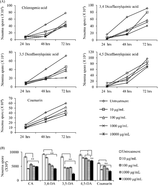
Colonies can be destroyed by nosemosis, which is brought on by a high Nosema spore infection in adult honey bees. The anti-nosemosis properties of ethanol and aqueous extracts from Aster scaber, Artemisia dubia, and their combination were previously documented. From these aqueous extracts, Balamurugan et al. separated five phenolic compounds (chlorogenic acid, 3,4-DCQA, 3,5-DCQA, 4,5-DCQA, and coumarin) and assessed their toxicities and anti-Nosema properties both in vitro and in vivo.
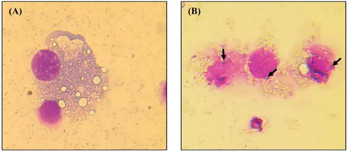
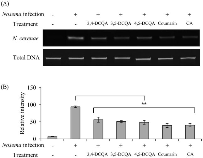
Nosema spores successfully infected IPL-LD-65Y cells (Fig. 1, black arrows). All substances showed anti-nosemosis action at different doses, according to in vitro screening (Fig. 2A). Interestingly, 10,000 μg/mL of coumarin and chlorogenic acid decreased spore counts to 40% and 42.3%, respectively, although 4,5-DCQA exhibited less action. Chlorogenic acid, coumarin, 3,4-DCQA, and 3,5-DCQA dramatically reduced spore populations following a 72-hour treatment (Fig. 2B). Nevertheless, toxicity rather than particular anti-parasitic activity was identified as the cause of the effects of 3,4-DCQA and 3,5-DCQA at maximal dose. Reduced N. ceranae DNA band intensities were confirmed by PCR analysis at 1000 μg/mL (Fig. 3A, B), with coumarin and chlorogenic acid showing the strongest anti-Nosema effects.
Ask a Question
Write your own review
Description: UMNSAH/DF-1 is a spontaneously immortalized chicken cell line derived from 10 day old East Lansing Line (ELL-0) eggs.

Description: Established from the larvae of the butterfly Mamestra brassicae (family Noctuidae, subfamily Noctuinae) (cabbage moth; Kohleule)
Description: This cell line is a subclone of the original cell line R1 established after elimination of mycoplasma with BM-Cyclin (tiamulin & minocycline)

Description: Derived from pupal ovarian tissue of spodoptera frugiperda. The cells are highly susceptible to Baculovirus infection and are used in the production of protein products genetically manipulated into ...

Description: Established from the embryonal (stage 52/53) normal heart endothelium (tadpole heart) of a South African clawed frog (Xenopus laevis Daudin) in 1979; cells were described to synthesize ...

Description: Established from the larvae of the Acrea Moth (Estigmene acrea; family Arctiidae, order Lepidoptera) (Bärenspinner)
- Adipose Tissue-Derived Stem Cells
- Human Neurons
- Mouse Probe
- Whole Chromosome Painting Probes
- Hepatic Cells
- Renal Cells
- In Vitro ADME Kits
- Tissue Microarray
- Tissue Blocks
- Tissue Sections
- FFPE Cell Pellet
- Probe
- Centromere Probes
- Telomere Probes
- Satellite Enumeration Probes
- Subtelomere Specific Probes
- Bacterial Probes
- ISH/FISH Probes
- Exosome Isolation Kit
- Human Adult Stem Cells
- Mouse Stem Cells
- iPSCs
- Mouse Embryonic Stem Cells
- iPSC Differentiation Kits
- Mesenchymal Stem Cells
- Immortalized Human Cells
- Immortalized Murine Cells
- Cell Immortalization Kit
- Adipose Cells
- Cardiac Cells
- Dermal Cells
- Epidermal Cells
- Peripheral Blood Mononuclear Cells
- Umbilical Cord Cells
- Monkey Primary Cells
- Mouse Primary Cells
- Breast Tumor Cells
- Colorectal Tumor Cells
- Esophageal Tumor Cells
- Lung Tumor Cells
- Leukemia/Lymphoma/Myeloma Cells
- Ovarian Tumor Cells
- Pancreatic Tumor Cells
- Mouse Tumor Cells