UMNSAH/DF-1
Cat.No.: CSC-C9259W
Species: Gallus gallus (Chicken)
Source: Embryo
Morphology: fibroblast
- Specification
- Background
- Scientific Data
- Q & A
- Customer Review
The UMNSAH/DF-1 cell line, originally isolated from 10-day-old East Lansing Line (ELL-0) chicken embryos, was found to be spontaneously transformed and free from any endogenous viral contamination. It exhibits high transfection efficiency and rapid proliferation, making it a commonly used chicken embryonic fibroblast line in research. DF-1 cells exhibit the characteristic spindle-shaped morphology typical of fibroblasts, featuring a uniform cytoplasm and distinct nucleoli. When densely packed, they develop into whorled or radial patterns. Despite their ability to form clones in semi-solid media, these cells have low tumorigenic potential, as they do not form tumors in immunocompromised mice. Additionally, they are susceptible to infections by avian poxvirus, avian reovirus, Marek's disease virus, and avian influenza virus, but notably, they do not harbor the ASLV genome associated with avian sarcoma viruses.
UMNSAH/DF-1 cells are widely used in various research fields, including:
Virus Proliferation and Recombinant Protein Expression: Researchers widely use this cell line for virus proliferation studies and recombinant protein expression and recombinant virus production because of its sensitivity to multiple viruses.
Drug Screening and Toxicology Studies: The UMNSAH/DF-1 cell line serves as an effective tool for drug toxicity evaluation because of its avian toxicity sensitivity.
Gene Cloning and Editing: These applications serve as an ideal model system for gene cloning procedures as well as gene editing techniques like CRISPR/Cas9 and gene expression research.
Biomaterial Evaluation: Research utilizes this system to determine how biomaterials interact biocompatibly with cells.
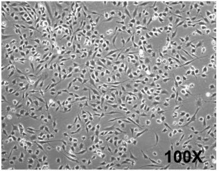 Fig. 1. Morphology and cell growth kinetics for DF-1 cells (Kong BW, Lee J Y, et al., 2011).
Enrofloxacin Induces Apoptosis and Reduces Viability of UMNSAH/DF-1 Cells
Enrofloxacin belongs to the fluoroquinolone category and has been used to treat bacterial infections in poultry since 1989. Current findings indicate that enrofloxacin potentially triggers apoptosis in multiple eukaryotic cell types which raises safety issues. Grabowski et al. examined its apoptotic effects on UMNSAH/DF-1 chicken cells, exploring the mitochondrial pathway as a potential mechanism. Methods include assessing changes in mitochondrial morphology, apoptosome formation, and caspase activation.
Researchers measured UMNSAH/DF-1, HEK-293, and PC3 cell viability using the MTT assay after treating them with enrofloxacin concentrations ranging from 2.5 to 40 μg/mL over time periods of 24 to 72 hours while comparing results to 10% DMSO (positive control) and medium (negative control). The experimental findings demonstrated that metabolic activity decreased over time in each tested cell line (Fig. 1). Further analysis with Annexin V and Dead Cell Assay at 10 μg/mL enrofloxacin showed increased apoptosis over time only in UMNSAH/DF-1 cells (Fig. 2A), corresponding to a typical veterinary dose for poultry. No significant apoptosis was noted in HEK-293 and PC3 lines, which retained a high number of live cells (Fig. 2B-C).
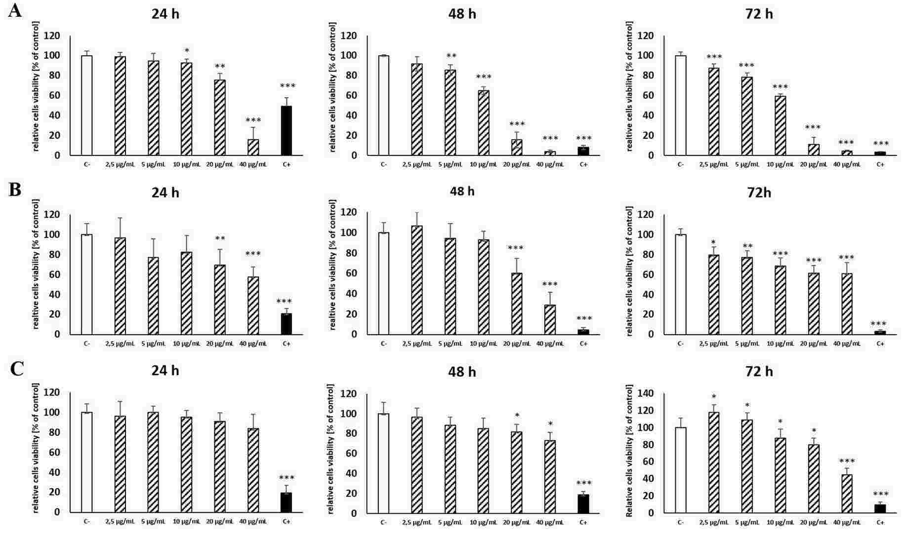 Fig. 1. Effects of enrofloxacin on viability of UMNSAH/DF-1 (panel A), HEK (panel B), and PC3 (panel C) cells after 24 h, 48 and 72 h of incubation, as assessed by the MTT test (Grabowski ?, Choszcz M, et al., 2024)
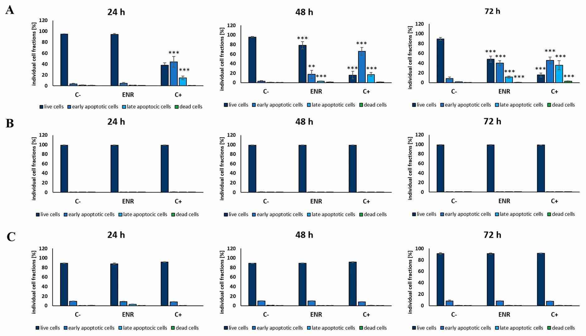 Fig. 2. Fractions of live, early-apoptotic, late-apoptotic and dead cells of the UMNSAH/DF-1 (panel A), HEK (panel B), and PC3 (panel C) cells after incubation in DMEM medium (negative control), DMEM with enrofloxacin (10 μg/mL), and DMEM with 10% DMSO (positive control) for 24 h, 48 h, 72 h (Grabowski ?, Choszcz M, et al., 2024).
PGPSt Alleviated Cr(VI)-Induced Mitochondrial Damage of DF-1 Cells
The heavy metal hexavalent chromium (Cr(VI)) has been acknowledged as a human carcinogen and both short-term and long-term exposure results in damage to organs. The compound PGPS originates from the Chinese herb Platycodon grandiflorus and displays multiple pharmacological properties. Therefore, Zhang's team investigated the role of PGPSt in Cr(VI)-induced apoptosis in chicken embryo fibroblast cell lines (DF-1 cells).
The roles of PGPSt in decreasing Cr(VI)-induced apoptosis in DF-1 cells were determined by analyzing oxidative stress and mitochondrial damage levels. The DF-1 cells underwent pretreatment with 150 μM Cr(VI) for a duration of 8 hours. Western blotting was used to examine Drp1 protein which plays an essential role in mitochondrial fission. As shown in Figure 3A, Drp1 expression increased significantly in the Cr(VI) group, indicating mitochondrial division. PGPSt treatment reduced Drp1 levels compared to the Cr(VI) group. Additionally, ROS, which activates Drp1, was measured. Figure 3B shows a significant ROS increase in the Cr(VI) group, reduced by PGPSt. MMP, an indicator of mitochondrial health, decreased in the Cr(VI) group but improved with PGPSt (Fig. 3C). These findings suggest Cr(VI) induces mitochondrial damage, while PGPSt mitigates it.
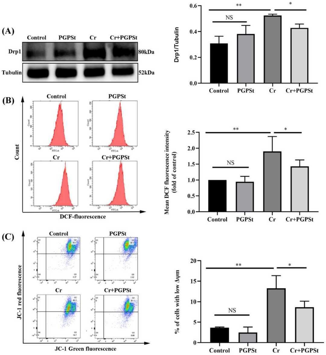 Fig. 3. PGPSt alleviated Cr(VI)-induced mitochondrial damage (Zhang Z, Zheng P, et al., 2022).
Ask a Question
Write your own review
- You May Also Need

Description: Established from the larvae of the butterfly Mamestra brassicae (family Noctuidae, subfamily Noctuinae) (cabbage moth; Kohleule)

Description: This cell line is a subclone of the original cell line R1 established after elimination of mycoplasma with BM-Cyclin (tiamulin & minocycline)

Description: Derived from pupal ovarian tissue of spodoptera frugiperda. The cells are highly susceptible to Baculovirus infection and are used in the production of protein products genetically manipulated into ...

Description: Established from the embryonal (stage 52/53) normal heart endothelium (tadpole heart) of a South African clawed frog (Xenopus laevis Daudin) in 1979; cells were described to synthesize ...

Description: Established from the larvae of the Acrea Moth (Estigmene acrea; family Arctiidae, order Lepidoptera) (Bärenspinner)
Description: Initiated from the choroid plexus of an ovine brain. The cells support propagation of Visna virus, strain 1514 with resulting cytopathic effects. Tests have indicated that the cell line produces both ...
- Adipose Tissue-Derived Stem Cells
- Human Neurons
- Mouse Probe
- Whole Chromosome Painting Probes
- Hepatic Cells
- Renal Cells
- In Vitro ADME Kits
- Tissue Microarray
- Tissue Blocks
- Tissue Sections
- FFPE Cell Pellet
- Probe
- Centromere Probes
- Telomere Probes
- Satellite Enumeration Probes
- Subtelomere Specific Probes
- Bacterial Probes
- ISH/FISH Probes
- Exosome Isolation Kit
- Human Adult Stem Cells
- Mouse Stem Cells
- iPSCs
- Mouse Embryonic Stem Cells
- iPSC Differentiation Kits
- Mesenchymal Stem Cells
- Immortalized Human Cells
- Immortalized Murine Cells
- Cell Immortalization Kit
- Adipose Cells
- Cardiac Cells
- Dermal Cells
- Epidermal Cells
- Peripheral Blood Mononuclear Cells
- Umbilical Cord Cells
- Monkey Primary Cells
- Mouse Primary Cells
- Breast Tumor Cells
- Colorectal Tumor Cells
- Esophageal Tumor Cells
- Lung Tumor Cells
- Leukemia/Lymphoma/Myeloma Cells
- Ovarian Tumor Cells
- Pancreatic Tumor Cells
- Mouse Tumor Cells