
KYSE170
Cat.No.: CSC-C6805J
Species: Homo sapiens (Human)
Source: Esophagus
Morphology: Epithelial-Like
- Specification
- Background
- Scientific Data
- Q & A
- Customer Review
The KYSE-170 cell line is a human esophageal squamous cell carcinoma (ESCC) model established from a primary tumor resected from a Japanese patient. As a primary tumor-derived line, KYSE-170 retains genetic and phenotypic features reflective of the clinical disease, making it a reliable and frequently utilized system in both basic and translational ESCC research.
KYSE-170 cells exhibit typical epithelial morphology and demonstrate aggressive growth properties consistent with carcinoma cells. Genomically, it is characterized by mutations commonly found in ESCC, including alterations in the TP53 tumor suppressor gene and CDKN2A (p16), contributing to dysregulated cell cycle progression and genomic instability. The line is tumorigenic in immunocompromised mice, forming solid tumors suitable for preclinical therapeutic studies. KYSE-170 has been extensively profiled at the genomic, transcriptomic, and proteomic levels, positioning it as one of the most well-annotated ESCC models available.
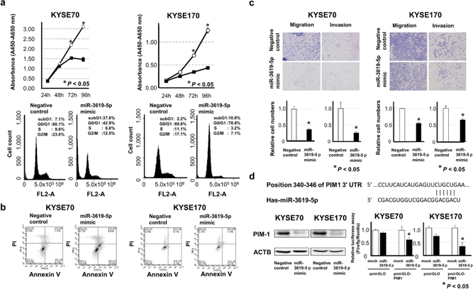
Low Levels of Tumor Suppressor miR-3619 Contribute to Malignant Outcomes and A Target for Nucleic Acid Therapy in Esophageal Cancer
Recent studies indicate that reduced levels of certain tumor-suppressing microRNAs (miRNAs) circulating in the blood are linked to tumor progression and poor prognosis across various types of malignancies. Identified from a comprehensive analysis of the NCBI and miRNA databases, we tested tumor suppressor miR-3619-5p in esophageal squamous cell carcinoma (ESCC). Both test-scale and large-scale analyses demonstrated that plasma levels of miR-3619-5p were markedly lower in ESCC patients than in healthy volunteers. Lower plasma levels of miR-3619-5p showed a strong association with advanced pathological stages and were recognized as an independent prognostic marker. Overexpression of miR-3619-5p in ESCC cells inhibited cell proliferation, migration and invasion through the direct suppression of novel target protein, proviral insertion site in Moloney murine leukemia virus 1 (PIM1). PIM1 is overexpressed in various solid and hematological cancers including ESCC, and has proven to be a promising target of inhibitors in recent clinical trials.
Ask a Question
Write your own review
- You May Also Need

Description: Human moderately differentiated squamous cell carcinoma cell line established from esophageal cancer.
Description: Human moderately differentiated squamous cell carcinoma cell line established from esophageal cancer.


Description: Human moderately differentiated squamous cell carcinoma cell line established from esophageal cancer.
Description: Human squamous cell carcinoma from oral cavity via mouse transplantation.

Description: Human squamous cell carcinoma cell line established from recurrent cancer (esophageal cancer).
- Adipose Tissue-Derived Stem Cells
- Human Neurons
- Mouse Probe
- Whole Chromosome Painting Probes
- Hepatic Cells
- Renal Cells
- In Vitro ADME Kits
- Tissue Microarray
- Tissue Blocks
- Tissue Sections
- FFPE Cell Pellet
- Probe
- Centromere Probes
- Telomere Probes
- Satellite Enumeration Probes
- Subtelomere Specific Probes
- Bacterial Probes
- ISH/FISH Probes
- Exosome Isolation Kit
- Human Adult Stem Cells
- Mouse Stem Cells
- iPSCs
- Mouse Embryonic Stem Cells
- iPSC Differentiation Kits
- Mesenchymal Stem Cells
- Immortalized Human Cells
- Immortalized Murine Cells
- Cell Immortalization Kit
- Adipose Cells
- Cardiac Cells
- Dermal Cells
- Epidermal Cells
- Peripheral Blood Mononuclear Cells
- Umbilical Cord Cells
- Monkey Primary Cells
- Mouse Primary Cells
- Breast Tumor Cells
- Colorectal Tumor Cells
- Esophageal Tumor Cells
- Lung Tumor Cells
- Leukemia/Lymphoma/Myeloma Cells
- Ovarian Tumor Cells
- Pancreatic Tumor Cells
- Mouse Tumor Cells