
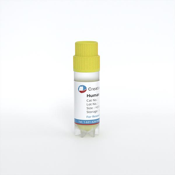
Human Corneal Endothelial Cells
Cat.No.: CSC-C2030WJ
Species: Human
Source: Cornea; Eye
Morphology: Polygonal
Cell Type: Endothelial Cell
- Specification
- Background
- Scientific Data
- Q & A
- Customer Review
Note: Never can cells be kept at -20°C.
Primary human corneal endothelial cells (HCECs) form the single layer of neuroectoderm-derived cells that lines the posterior surface of the cornea. In vivo, these post-mitotic cells are essential for maintaining corneal transparency through a “pump-leak” mechanism. Primary HCECs, isolated from donor tissue and cultured under optimized conditions, recapitulate this specialized physiology in vitro, providing a faithful human model of the corneal endothelium.
A key strength of primary HCECs is their capacity to establish a fully differentiated, contact-inhibited monolayer with characteristic polygonal, often hexagonal, morphology. They organize robust apicolateral tight junctions, visualized by continuous ZO-1 localization, and uniformly express the defining endothelial markers Na⁺/K⁺-ATPase, aquaporin-1, N-cadherin, and collagen VIII, while remaining negative for corneal epithelial keratins. Functionally, these cells generate a measurable transendothelial electrical resistance and actively transport ions and fluid, thereby mirroring the deturgescence pump critical for corneal clarity. Their human provenance eliminates species-dependent confounders, enabling direct investigation of endothelial dystrophies such as Fuchs endothelial corneal dystrophy, assessment of drug-induced toxicity, and development of tissue-engineered grafts. Notably, supplementation with Rho-associated kinase (ROCK) inhibitors now permits transient expansion of primary HCECs without sacrificing differentiated function, overcoming their inherently limited proliferative capacity and opening avenues for cell-based regenerative therapy.
AMF30a Promotes the Proliferation and Adhesion of Human Corneal Endothelial Cells
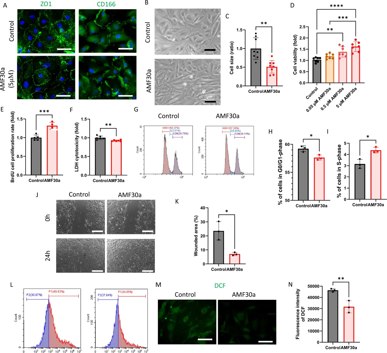
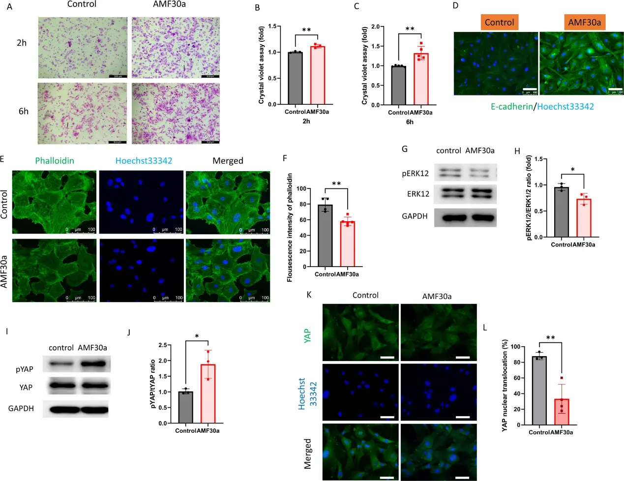
Corneal endothelial cells (CECs), located in the innermost layer of cornea, are crucial for maintaining its transparency. Peptidylarginine deiminase 2 (PAD2) is an enzyme responsible for catalyzing the post-translational modification of arginine into citrulline, a process known as citrullination. This study investigated the effect of AMF30a, PAD2 inhibitor, on survival and function of CECs.
Cultured human CECs (hCECs) were treated with AMF30a, and analyzed for viability (CCK-8), proliferation (BrdU assay), cytotoxicity (LDH assay), and oxidative stress (DCF-DA). Adhesion and morphology were evaluated via crystal violet staining and imaging software. The results revealed that AMF30a enhanced cell viability, proliferation, and adhesion while reducing cytotoxicity and oxidative stress. Morphological analysis revealed reduced cell size and elongation factor, indicating structural changes.
Ask a Question
Write your own review
Description: Human Iris Pigment Epithelial Cells (HIPEpiC) from Creative Bioarray are isolated from the human iris. HIPEpiC are cryopreserved at passage one and delivered frozen. Each vial contains >5 x 10^5 ...
Description: Human choroidal endothelial cells are isolated from normal human choroidal tissue. The cells are provided at passage 1 and shipped in frozen vials. These cells have a minimum average population ...
Description: Human Conjunctival Epithelial Cells (HConEC) from Creative Bioarray are isolated from human conjunctiva. HConEC are cryopreserved from passage one culture. Each vial contains >5 x 10^5 cells in 1 ml ...
Description: Human Trabecular Meshwork Cells (HTMC) are isolated from the juxtacanalicular and corneoscleral regions of human eye. HTMC are cryopreserved at P0 and delivered frozen. Each vial contains >5 x 10^5 ...
Description: Human Non-Pigment Ciliary Epithelial Cells (HNPCEpiC) from Creative Bioarray are isolated from human ciliary bodies. HNPCEC are cryopreserved at primary culture and delivered frozen. Each vial ...
Description: Human Retinal Astrocytes (HRA) from Creative Bioarray are isolated from human retina. HRA are cryopreserved at passage one and delivered frozen. Each vial contains >5 x 10^5 cells in 1 ml volume. HRA ...