
Advanced Organoid Culture: A Practical Optimization Guide
You are staring at your third batch of intestinal organoids and something is wrong. The first batch grew into beautiful budding structures. The second batch partially collapsed. This one barely grew at all. You have scanned the literature, but what you really need is a clear, actionable framework: when organoid culture fails, where do you start?
This guide is designed to answer that question. We will not recite every paper ever published. Instead, we will walk through a structured decision-making process you can apply immediately, whether you are growing intestinal, brain, kidney, liver, or tumor organoids. The principles are universal, even if the details vary.
The Optimization Framework: A Systematic Approach
When organoids are not behaving, most people reflexively tweak growth factors. That is usually the wrong place to start. Here is a better sequence:
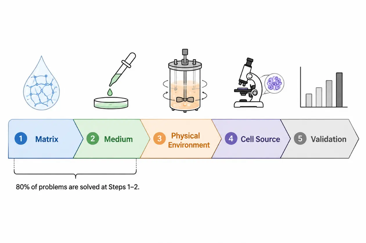
This order is intentional. Extracellular matrix is the single most influential variable in organoid culture, yet it receives the least systematic attention in most labs. Master the matrix, and half your problems disappear.
Step 1: Matrix Selection — Your First and Most Important Decision
Ask yourself three questions before touching anything else:
1. What is my endpoint?
If you are publishing a mechanistic study, continuing with Matrigel is acceptable—provided you record the lot number
and acknowledge its limitations. If you are doing drug screening, toxicity testing, or preparing for clinical
translation, you must switch to a defined matrix now.
2. Is my organoid type sensitive to matrix stiffness?
Brain organoids prefer soft matrices (~0.1–0.5 kPa), while intestinal organoids tolerate stiffer environments (~1–3
kPa). If your organoids are collapsing or failing to expand, the stiffness may simply be wrong for your tissue.
3. Am I using the right adhesion cues?
Matrigel is rich in laminin and collagen IV. If you switch to a synthetic matrix, you must supply the appropriate
adhesion ligands. RGD peptides work for many epithelial types, but neural organoids often require laminin-derived
peptides like IKVAV or YIGSR.
Choosing a practical alternative
If you decide to move beyond Matrigel, here is a decision tree:
- For intestinal organoid expansion: Start with PEG-4MAL hydrogels. The protocol by Cruz-Acuña et al. (2018) is detailed and reproducible. You will need 4-arm PEG-maleimide, RGD peptide, and a protease-degradable crosslinker peptide. Mix at room temperature; gelation occurs within 15 minutes. Total cost per 96-well plate is roughly comparable to growth-factor-reduced Matrigel.
- For brain organoids: Chemically defined hyaluronic acid hydrogels offer a rapid induction protocol. The Lindborg et al. (2016) approach shortens culture time to about four weeks and eliminates the undefined variables that make brain organoid protocols notoriously inconsistent.
- For kidney organoids: Soft matrices (~0.5 kPa) accelerate nephron formation. Garreta et al. (2019) showed that tuning substrate stiffness modulates YAP signaling, which directly influences kidney progenitor differentiation. If your kidney organoids are slow to mature, try softening your matrix first.
- For cost-sensitive labs: Tissue-derived decellularized ECM hydrogels from porcine intestinal submucosa are a practical option. Kim et al. (2022) demonstrated that these matrices support gastrointestinal organoid growth at approximately one-fifth the cost of Matrigel. The trade-off is more hands-on preparation time.
Practical matrix checklist
Use this checklist every time you prepare a new batch of matrix:
- Record the lot number (even for Matrigel)
- Measure or verify stiffness if possible (AFM or rheometry)
- Test at least two RGD peptide concentrations (.5 mM and 2 mM) when using synthetic matrices
- Verify that the degradation rate matches organoid growth rate
- If using synthetic matrix, confirm that organ-specific growth factors are added
- Pre-warm all reagents to 4°C before mixing Matrigel to prevent premature polymerization
Step 2: Medium Optimization — Less Is Often More
The most common mistake in organoid culture is growth factor over-supplementation. Researchers read a protocol, add a few extra factors for good measure, and end up with undifferentiated cystic structures that refuse to mature.
Fujii et al. (2018) demonstrated this elegantly in intestinal organoids. Simplifying the standard "Wnt + EGF + Noggin + R-spondin" cocktail to "low Wnt + EGF + Noggin" dramatically increased Paneth cell and enteroendocrine cell differentiation while maintaining stem cell self-renewal. The principle extends across organoid types: more factors do not equal better culture.
A systematic approach to medium optimization
Phase 1: Identify the minimal essential factors. Run a factor withdrawal experiment. Remove one factor at a time and observe the effect over two passages. You will typically find that only two or three factors are absolutely essential. Everything else is either supportive or superfluous.
Phase 2: Titrate the critical factors. Wnt concentration is the most powerful lever for controlling the self-renewal versus differentiation balance in epithelial organoids. Start with a three-point titration (0.5×, 1×, 2× the standard concentration) and evaluate morphology and marker expression. BMP inhibitors (Noggin, GREM1) are the second most important lever: too much inhibition blocks differentiation entirely.
Phase 3: Add tissue-specific factors. Once the basal medium is optimized, you can add factors that promote specific cell types. For example, IL-22 promotes Paneth cell formation in intestinal organoids (He et al., 2022). Hypoxia conditions promote airway differentiation in lung organoids (Dong et al., 2025). For liver organoids, removing Wnt and adding dexamethasone promotes hepatocyte maturation.
Conditioned medium versus recombinant proteins
This is a practical trade-off every lab faces:
| Conditioned Medium | Recombinant Proteins | |
|---|---|---|
| Cost | Low | High |
| Batch consistency | Poor | Excellent |
| Best for | Early-stage exploration | Optimization and standardization |
| Practical advice | Use initially, but prepare to switch | Stock up for critical experiments |
If you use conditioned medium, dedicate time to characterizing each batch. A simple activity assay using a Wnt or BMP reporter cell line can save you months of troubleshooting downstream.
Step 3: Physical Environment — The Overlooked Variable
Most labs culture organoids in standard CO₂ incubators at atmospheric oxygen (~20% O₂). But most tissues in the body exist at 1–8% O₂. This discrepancy matters.
- Oxygen tension. Intestinal organoids expand better at 5% O₂. Brain organoids develop oxygen gradients naturally, with central regions becoming hypoxic and necrotic if the organoid grows too large. If you see central necrosis, reduce oxygen concentration or switch to a spinning bioreactor to improve diffusion.
- Matrix geometry. This is a recent and powerful insight: the initial shape of the matrix droplet determines organoid patterning. Gjorevski et al. (2022) showed that seeding intestinal stem cells in tubular molds promotes crypt-villus axis formation. Hemispherical droplets promote cortical folding in brain organoids. Microwell plates produce more uniformly sized organoids. If you are struggling with reproducibility, try constraining the initial geometry.
- Mechanical forces. Spinning bioreactors improve nutrient diffusion. Cyclic stretch promotes alveolar organoid maturation. Hydrostatic pressure affects kidney tubule formation. The principle is simple: if your organoid normally experiences mechanical forces in vivo, it probably needs them in vitro.
Step 4: Reproducibility — The Foundation of Everything
| Problem | Likely Cause | Solution |
|---|---|---|
| Variable organoid size | Inconsistent seeding density | Use an automated cell counter; seed at a fixed density per well |
| Organoid collapse or rupture | Matrix degradation too fast | Increase crosslinking density or add a protease inhibitor cocktail |
| Central necrosis | Nutrient or oxygen diffusion limit | Reduce organoid size, switch to spinning culture, or lower oxygen |
| Poor differentiation | Wnt/BMP signaling imbalance | Reduce Wnt concentration; add BMP or remove BMP inhibitor |
| Low post-passage survival | Single-cell dissociation stress | Add ROCK inhibitor Y-27632 at 10 μM for the first 24 hours after passaging |
| Batch-to-batch variability | Undefined matrix components | Reserve a single large Matrigel lot for critical experiments, or switch to a defined matrix |
| Contamination | Mycoplasma | Test monthly; discard positive cultures immediately |
You cannot optimize what you cannot measure, and you cannot reproduce what you do not document. Establish these minimum standards:
- Mycoplasma testing every month
- Freeze at least three vials of every organoid line as backup
- Record the lot number and preparation date of every medium batch
- Validate cell type composition regularly (at minimum, qPCR for key markers; ideally, periodic scRNA-seq)
- Photograph organoids at every passage to build a morphological reference atlas
Establish an Internal Gold Standard
Select one well-characterized organoid line as your laboratory's positive control. Include it in every new batch of culture. If the control line fails, the problem is environmental—matrix, medium, or equipment—not your experimental line. This single practice will save you more time than any other optimization strategy.
When You Are Ready to Expand: Vascularization and Co-Culture
Once your organoid culture is stable and reproducible, adding complexity becomes feasible.
Vascularization has two practical paths. The simpler route is in vivo transplantation: implant organoids under the kidney capsule of immunodeficient mice, and host blood vessels will infiltrate within two to four weeks. This works for terminal experiments but is not suitable for screening. The in vitro route involves co-culturing organoids with endothelial cells (HUVEC or iPSC-derived) in a fibrin gel matrix. Add VEGF at 50 ng/mL and FGF2 at 25 ng/mL; expect capillary-like networks within seven to ten days.
Immune co-culture requires a key decision: autologous immune cells from the same donor, or allogeneic PBMCs from healthy donors? Autologous cells better reflect individual immune responses but are limited in supply. Allogeneic cells are convenient for initial screening. A typical T cell killing assay uses a 5:1 to 10:1 effector-to-target ratio, with 100 U/mL IL-2 to maintain T cell viability. Readouts are typically collected at 48–72 hours.
Key Principles
- Optimize matrix first, then medium, then physical environment. This sequence maximizes efficiency.
- Document everything. Lot numbers, concentrations, observations—these data are the foundation of optimization.
- Simplify before you complexify. Verify that every factor in your medium is truly necessary.
- Always run a positive control. Without one, you are optimizing in the dark.
- Let your organoids tell you what they need. The most reliable optimization signal comes from the cells themselves, not from the latest paper.
Creative Bioarray Relevant Recommendations
| Products & Services | Description |
|---|---|
| 3D Spheroid Platform for Drug Development | Creative Bioarray provides advanced 3D spheroid models designed for researchers aiming to explore cell behavior in a more realistic environment. |
| Organoid Platform for Drug Development | Creative Bioarray presents a state-of-the-art Organoid Platform designed to transform drug development and biomedical research. By leveraging advanced organoid and organoid-immune co-culture models, this platform provides highly accurate and predictive solutions that closely replicate human biology. |
| Organ-on-a-Chip Platform for Drug Development | Creative Bioarray focuses on developing specific human disease models and leveraging our organ-on-a-chip platform to identify new therapeutics and clinical biomarkers, promote drug development, and create novel drug delivery systems. |