
Human Trabecular Meshwork Cells
Cat.No.: CSC-C1535
Species: Human
Source: Trabecular Meshwork; Eye
Cell Type: Trabecular Meshwork Cell
- Specification
- Background
- Scientific Data
- Q & A
- Customer Review
Human Trabecular Meshwork Cells (HTMCs) are primary mesenchymal-derived cells that line the collagenous beams of the trabecular meshwork, the principal tissue regulating aqueous humor outflow resistance and intraocular pressure (IOP). As non-immortalized diploid cells, they faithfully preserve native phagocytic function, contractile properties, and the extracellular matrix (ECM) remodeling machinery essential for maintaining outflow facility.
A paramount advantage of HTMCs is their unique capacity to recapitulate the pathogenic cascades of primary open-angle glaucoma (POAG) and steroid-induced glaucoma in vitro. Upon stimulation with TGF-β2, the central glaucomatous cytokine, HTMCs robustly undergo myofibroblastic differentiation, marked by α-smooth muscle actin expression, formation of cross-linked actin networks, and excessive deposition of fibronectin and collagens, thereby directly modeling the outflow resistance characteristic of POAG. Moreover, HTMCs exhibit exquisite glucocorticoid sensitivity, responding to dexamethasone with pronounced induction of the glaucoma-associated protein myocilin and ECM accumulation, faithfully replicating the steroid-induced glaucomatous phenotype. They also display senescence-associated secretory phenotype and oxidative stress responses that drive progressive dysfunction, enabling dissection of age-related IOP elevation.
HTMCs form confluent monolayers on Transwell inserts, permitting quantitative measurement of hydraulic conductivity and pharmacological evaluation of outflow-facilitating agents such as ROCK inhibitors and nitric oxide donors. Collectively, HTMCs represent the indispensable human platform for investigating aqueous humor dynamics, glaucoma pathogenesis, and preclinical screening of IOP-lowering therapeutics.
Metformin Alleviates Fibrosis of Trabecular Meshwork Cells Induced by TGFβ2
In this study, Ren, Jing, et al. investigated the protective effect of Metformin on fibrosis of trabecular meshwork cells induced by TGFβ2.
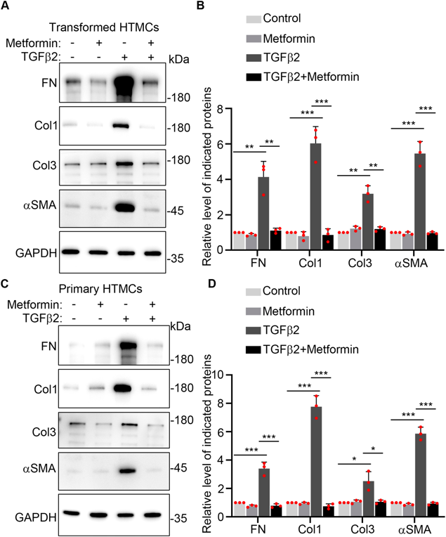
They found 400 μM Metformin activates autophagy in human trabecular meshwork cells (HTMCs), and autophagy activation could ameliorate fibrosis, so they wonder whether Metformin could alleviate fibrosis of HTMCs. TGFβ2 was usually used to induce fibrosis of HTMCs in vitro, and they treated transformed and primary HTMCs with 400 μM Metformin, 10 ng/mL TGFβ2, or 400 μM Metformin+10 ng/mL TGFβ2 for 48 h. Western blotting data showed fibrotic proteins levels were significantly up-regulated after TGFβ2 treatment, while 400 μM Metformin co-treatment significantly decreased fibrotic proteins expression levels compared with TGFβ2 treatment. 400 μM Metformin treatment alone didn't affect fibrotic proteins expression levels (Fig. 1A-D).
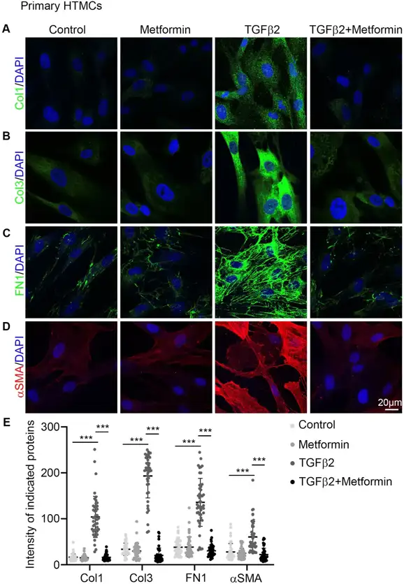
To confirm whether 400 μM Metformin could reduce fibrosis of HTMCs induced by TGFβ2, they used immunofluorescence staining assay to detect FN, Col1, Col3 and αSMA levels in primary HTMCs. Confocal images and fluorescence quantification data showed that TGFβ2 strongly increased FN, Col1, Col3 and αSMA fluorescence intensities, while 400 μM Metformin co-treatment significantly repressed fibrotic proteins fluorescence intensities compared with TGFβ2 treatment alone (Fig. 2A-E).
Ask a Question
Write your own review
Description: Human choroidal endothelial cells are isolated from normal human choroidal tissue. The cells are provided at passage 1 and shipped in frozen vials. These cells have a minimum average population ...
Description: Human Non-Pigment Ciliary Epithelial Cells (HNPCEpiC) from Creative Bioarray are isolated from human ciliary bodies. HNPCEC are cryopreserved at primary culture and delivered frozen. Each vial ...
Description: Human Keratocytes (HK) from Creative Bioarray are isolated from human cornea. HK are cryopreserved at primary culture. Each vial contains >5 x 10^5 cells in 1 ml volume. HK are characterized by their ...
Description: RFP Expressing Human Retinal Microvascular Pericytes (RFP-HRMPs) provided by Creative Bioarray are zeocin-selected from Human Retinal Microvascular Pericytes infected with lentivirus expressing RFP. ...
Description: Primary Human Retinal Pericyte Cells (HRPEpiC) are isolated from human retina. HRPEpiC are cryopreserved at passage one and delivered frozen. Each vial contains >5 x 10^5 cells in 1 ml volume. ...
Description: Human Retinal Pigment Epithelial Cells (HRPEpiC) are isolated from human retina. HRPEpiC are cryopreserved at passage one and delivered frozen. Each vial contains >5 x 10^5 cells in 1 ml volume. ...