
Human Conjunctival Epithelial Cells (HConEC)
Cat.No.: CSC-7802W
Species: Human
Source: Conjunctiva; Eye
Cell Type: Epithelial Cell
- Specification
- Background
- Scientific Data
- Q & A
- Customer Review
Human conjunctival epithelial cells (HCECs) form a non-keratinized, stratified epithelium that lines the inner surface of the eyelids and covers the anterior sclera, interspersed with mucin-secreting goblet cells. Together with the corneal epithelium, they maintain ocular surface integrity by providing a physical barrier, producing tear film-stabilizing mucins, and participating in innate immune surveillance. Primary HCECs, isolated from donor tissue and cultured under optimized conditions, faithfully retain native phenotypic characteristics, offering a physiologically relevant human in vitro model.
A defining advantage of primary HCECs is their capacity to differentiate into a stratified, polarized epithelium with functional tight junctions, establishing a competent permeability barrier. Critically, when cultured at the air–liquid interface, they generate goblet cells that secrete the gel-forming mucin MUC5AC, alongside membrane-tethered mucins MUC1 and MUC16, thereby recapitulating the conjunctival glycocalyx–tear film interface. The cells constitutively express conjunctival keratins (K4, K13, K19) and a repertoire of pattern recognition receptors (TLR2, TLR3, TLR4) and histamine receptors, enabling mechanistic investigation of allergic and microbial responses. In contrast to immortalized cell lines, which frequently exhibit genomic instability and loss of goblet cell differentiation, primary HCECs maintain diploid genomes, donor-specific features, and robust regulated mucin secretion. Their human origin eliminates species-dependent confounders, strengthening translational relevance for prevalent ocular surface disorders including dry eye disease, allergic conjunctivitis, and chemical injury.
Characteristics of X-irradiated Corneal and Conjunctival Epithelial Cells
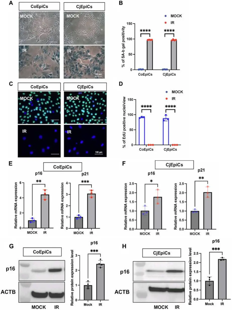
Elucidating the role of senescent ocular surface cells could increase our knowledge of the pathology of refractory age-related ocular surface diseases, including dry eye and limbal stem cell deficiency (LSCD). Here, we induced cellular senescence in human corneal epithelial cells (CoEpiCs) and conjunctival epithelial cells (CjEpiCs) using X-irradiation, and analyzed gene expression profiles of each cell type to determine the characteristics of senescent ocular surface cells.
As controls (mock), non-irradiated CoEpiCs and CjEpiCs showed homogenous populations of small cells. However, X-irradiated CoEpiCs and CjEpiCs appeared more elongated and flattened, and significantly bigger than non-irradiated cells (Fig. 1A). SA-β-gal in irradiated CoEpiCs and CjEpiCs was revealed in blue (Fig. 1B). EdU-labeling was not detectable in irradiated CoEpiCs and CjEpiCs, whereas 91.3% and 88.3 % of non-irradiated CoEpiCs and CjEpiCs were EdU-positive (Fig. 1C, 1D). We next examined the expression of the senescence markers p16 and p21. Using qRT-PCR, both p16 and p21 were significantly elevated in irradiated CoEpiCs and CjEpiCs compared to non-irradiated cells (Fig. 1E, 1F), and western blotting for p16 expression showed similar differences between cell populations (Fig. 1G, 1H), suggesting that irradiated CoEpiCs (SCo) and CjEpiCs (SCj) are indeed senescent.
Ask a Question
Write your own review
Description: Human choroidal endothelial cells are isolated from normal human choroidal tissue. The cells are provided at passage 1 and shipped in frozen vials. These cells have a minimum average population ...
Description: Human Non-Pigment Ciliary Epithelial Cells (HNPCEpiC) from Creative Bioarray are isolated from human ciliary bodies. HNPCEC are cryopreserved at primary culture and delivered frozen. Each vial ...
Description: Human Keratocytes (HK) from Creative Bioarray are isolated from human cornea. HK are cryopreserved at primary culture. Each vial contains >5 x 10^5 cells in 1 ml volume. HK are characterized by their ...

Description: RFP Expressing Human Retinal Microvascular Pericytes (RFP-HRMPs) provided by Creative Bioarray are zeocin-selected from Human Retinal Microvascular Pericytes infected with lentivirus expressing RFP. ...

Description: Primary Human Retinal Pericyte Cells (HRPEpiC) are isolated from human retina. HRPEpiC are cryopreserved at passage one and delivered frozen. Each vial contains >5 x 10^5 cells in 1 ml volume. ...
Description: Human Retinal Pigment Epithelial Cells (HRPEpiC) are isolated from human retina. HRPEpiC are cryopreserved at passage one and delivered frozen. Each vial contains >5 x 10^5 cells in 1 ml volume. ...