
ddPCR Assay Design for Gene Rearrangement Detection
Gene Rearrangements Suitable for ddPCR Detection
Gene rearrangements are structural abnormalities of the genome that are widely used as diagnostic and therapeutic biomarkers in cancer research. Common forms include gene fusions, chromosomal translocations and inversion-derived rearrangements many of which drive tumor development or progression.
Because droplet digital PCR (ddPCR) is a targeted detection technology, it is particularly effective for analyzing known gene rearrangements with defined breakpoint sequences or fusion transcripts. High sensitivity and absolute quantification make it ideally suited for biomarker validation, monitoring minimal residual disease (MRD) and confirmation of sequencing results.
Some representative examples are summarized below.
| Gene Rearrangement | Disease Association | Sample Type |
|---|---|---|
| BCR::ABL1 | Chronic myeloid leukemia | Blood, bone marrow |
| EML4::ALK | Non-small cell lung cancer | FFPE tissue |
| PML::RARA | Acute promyelocytic leukemia | Bone marrow |
| TMPRSS2::ERG | Prostate cancer | Tissue biopsy |
These well-characterized rearrangements provide ideal targets for customized ddPCR assay development.
Selecting the Appropriate Assay Strategy
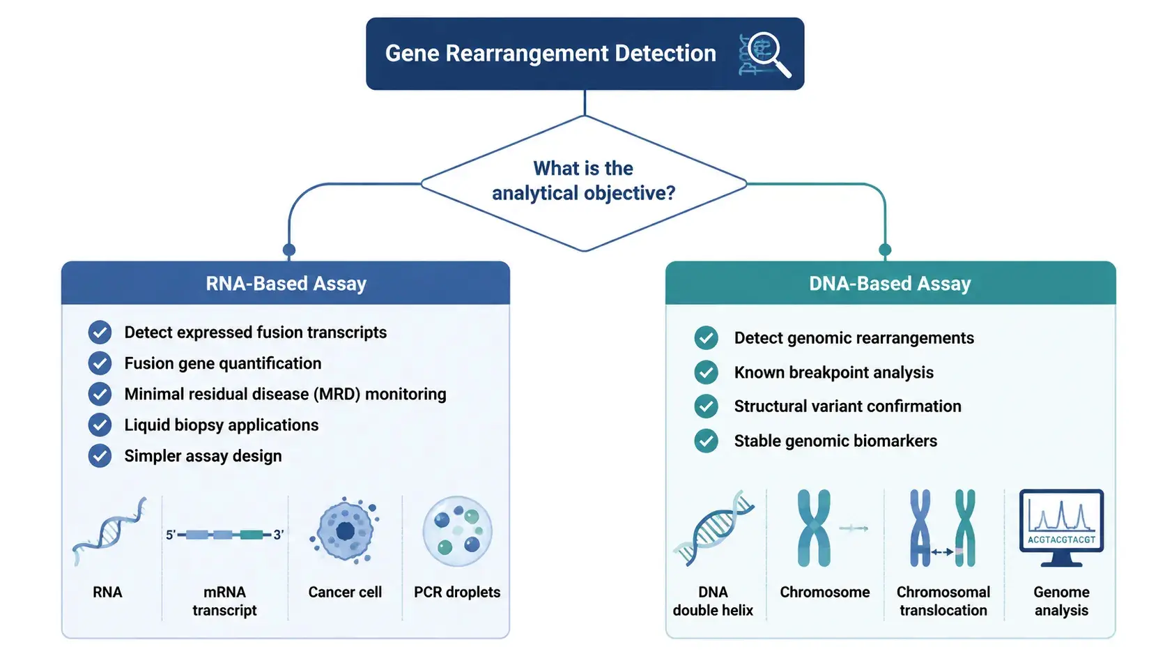
The first step in reliable detection of gene rearrangements is to select the proper test technique. The choice depends mostly on the biological features of the target and on the type of accessible sample.
- RNA-based assays are preferred for detecting expressed fusion transcripts because they directly measure the functional products of gene rearrangements. These are commonly employed for measurement of fusion genes, therapy monitoring and MRD research. Because intronic sequences are spliced out during RNA splicing, assay design is usually simpler.
- DNA-based assays target genomic breakpoints and are useful when the breakpoint sequence is known or when genomic alterations rather than transcript expression are of interest. However, large intronic regions and breakpoint heterogeneity often make assay design more challenging.
In general, RNA is the preferred template for most fusion gene assays, while DNA-based detection is more suitable for selected structural rearrangement studies.
Designing a Reliable ddPCR Assay
After selecting the proper assay approach, careful design of primers and probes is the most important aspect affecting test specificity and sensitivity. Unlike conventional PCR, ddPCR partitions each reaction into thousands of nanoliter-sized droplets, which makes the test performance extremely dependent on effective amplification and clear discrimination between positive and negative droplets.
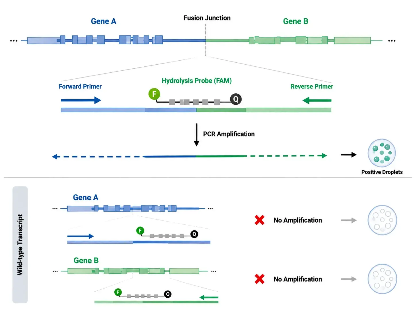
For RNA-based fusion detection, primers should be designed to flank the fusion junction, while the hydrolysis probe spans the breakpoint whenever possible. This configuration minimizes amplification of wild-type transcripts and ensures that only the target fusion transcript generates fluorescent signals.
For DNA-based assays, primer placement depends on the genomic breakpoint. Because breakpoints often occur within large intronic regions and may vary among patients, DNA assays are generally feasible only when breakpoint sequences have been previously characterized. When breakpoint heterogeneity exists, multiple primer/probe sets may be required to achieve comprehensive detection.
Several general design principles should also be considered:
| Design Parameter | Recommended Practice |
|---|---|
| Primer length | 18–24 nt |
| GC content | 40–60% |
| Amplicon size | 60–120 bp (preferably <100 bp for FFPE samples) |
| Probe Tm | 68–70°C (approximately 8–10°C higher than primers) |
| Primer Tm | 58–62°C |
| Secondary structures | Avoid hairpins, dimers, and long homopolymers |
| Fusion junction | Probe preferably overlaps the breakpoint |
Hydrolysis probes labelled with distinct fluorophores (e.g., FAM and HEX/VIC) enable multiplex detection of fusion targets along with internal reference genes in the same experiment. Probe placement directly across the fusion junction usually provides the highest analytical specificity because fluorescence is generated only when the exact fusion transcript is present.
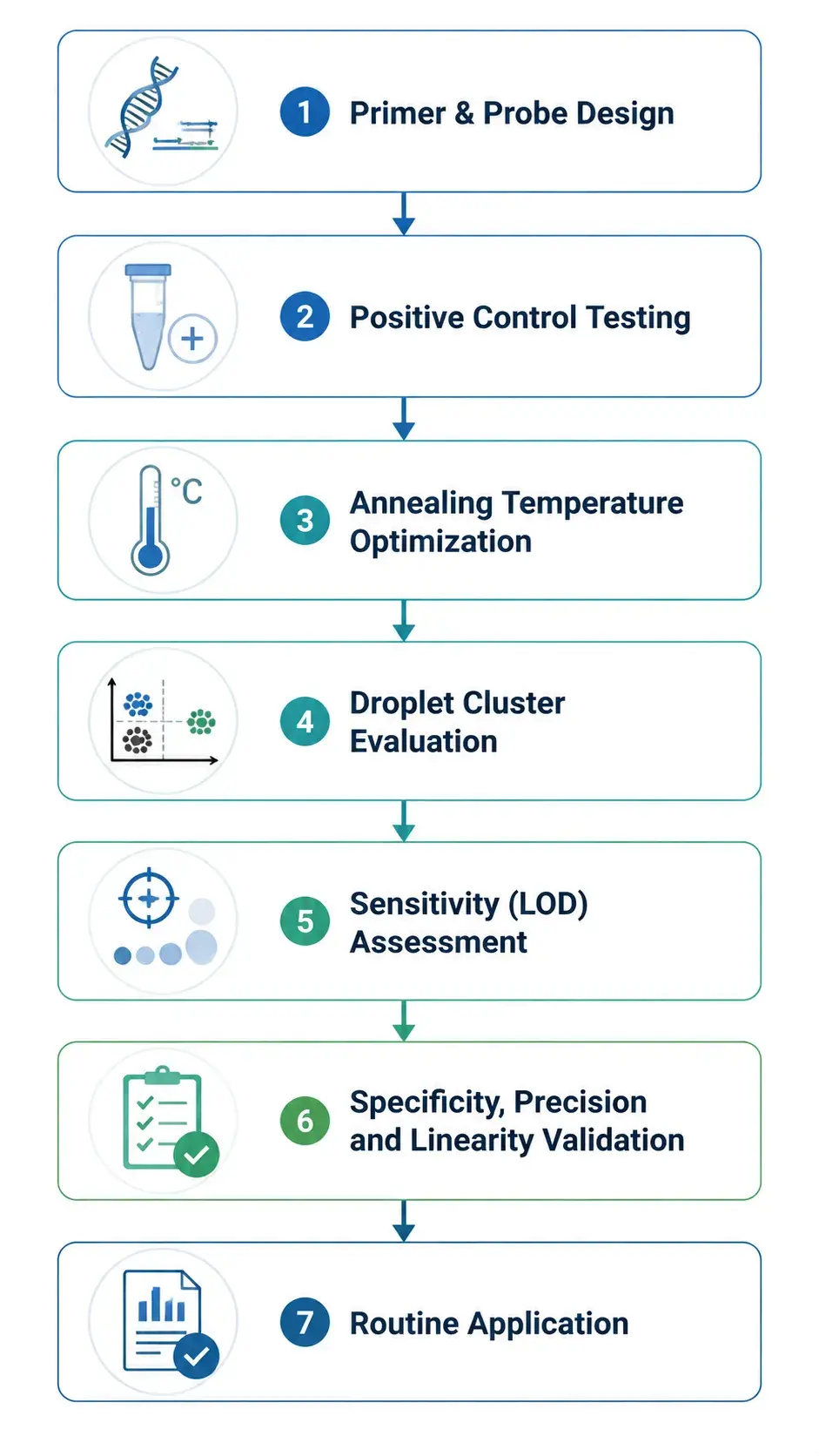
Once the primer and probe sequences are constructed, preliminary optimization with gradient annealing temperatures and positive control materials is required. Well-separated droplet clusters with little "rain" indicate satisfactory assay performance and give confidence for the subsequent analytical validation.
Assay Validation Before Routine Testing
Before applying a ddPCR assay to routine research or translational studies, its analytical performance should be thoroughly validated to assure reliable and reproducible results. Systematic validation method helps to build trust in assay sensitivity, specificity and quantitative accuracy
Key validation parameters include:
- Analytical Sensitivity: Determine the lowest detectable target concentration by testing serial dilutions of positive control material. This establishes the assay's limit of detection (LOD).
- Analytical Specificity: Confirm that the assay detects only the intended fusion target without amplifying wild-type sequences or unrelated rearrangements.
- Precision: Assess repeatability by analyzing technical replicates within the same experiment and across independent runs.
- Linearity: Evaluate quantitative performance over a range of target concentrations to ensure consistent and proportional measurement.
- Robustness: Verify assay performance under minor experimental variations, such as different reagent lots, operators, or instruments.
| Validation Parameter | Purpose |
|---|---|
| Analytical Sensitivity | Determine the limit of detection |
| Analytical Specificity | Confirm target-specific amplification |
| Precision | Evaluate repeatability across replicates |
| Linearity | Verify quantitative performance |
| Robustness | Assess assay consistency under routine conditions |
A well-validated assay provides a solid foundation for downstream applications and minimizes variability between experiments.
Best Practices for Gene Rearrangement Detection
Careful planning and standardized experimental procedures are essential for developing reliable ddPCR assays. The following recommendations can help improve assay performance and reduce optimization time.
- Confirm the fusion sequence and transcript variant before designing primers and probes.
- Design primers that specifically span the fusion junction to maximize assay specificity.
- Select hydrolysis probes with appropriate fluorophore and quencher combinations for clear signal detection.
- Use experimentally validated reference genes when quantitative normalization is required.
- Include fusion-positive, wild-type, and no-template controls in every assay.
- Optimize annealing temperature and reaction conditions before large-scale experiments.
- Validate assay performance using dilution series and independent replicate experiments.
- Document assay parameters and validation results to support long-term reproducibility.
Applications of ddPCR in Gene Rearrangement Analysis
With high sensitivity and absolute quantification capability, ddPCR has become a valuable tool for a wide range of gene rearrangement studies.
One of its primary applications is oncology biomarker research, where known fusion genes such as BCR::ABL1, EML4::ALK, and RET fusions are quantified to investigate disease biology and evaluate targeted therapies.
ddPCR is also widely used for minimal residual disease (MRD) monitoring. Its ability to detect extremely low levels of fusion transcripts makes it suitable for tracking treatment response and identifying molecular relapse during long-term follow-up.
Another important application is orthogonal validation of sequencing results. Candidate fusion events identified by RNA sequencing or targeted NGS panels can be rapidly confirmed and accurately quantified using customized ddPCR assays.
In translational research, ddPCR supports biomarker verification, analytical assay development, and companion diagnostic studies by providing sensitive and reproducible quantitative measurements.
| Application | Typical Purpose |
|---|---|
| Fusion gene quantification | Biomarker research |
| MRD monitoring | Treatment response evaluation |
| NGS result confirmation | Orthogonal validation |
| Biomarker verification | Translational research |
| Assay development | Method optimization and validation |
Conclusion
Droplet digital PCR is a powerful platform for the sensitive detection and absolute quantification of known gene rearrangements. However, its effectiveness is highly dependent on thorough assay creation, including suitable target selection, optimized primer and probe design, and rigorous analytical validation.
By applying proven assay design concepts and standardized validation processes, researchers can provide highly repeatable data for fusion gene analysis, biomarker verification, and disease monitoring. As precision medicine is still in progress, ddPCR continues to be a valid and complementary tool for gene rearrangement research, especially where precise quantification of known fusion events is needed.
Creative Bioarray Relevant Recommendations
| Products & Services | Description |
|---|---|
| Droplet Digital PCR (ddPCR) Service | Our experienced team at Creative Bioarray provides expert ddPCR analysis support with advanced technical capabilities and customized solutions for diverse molecular research applications. |