
Rat Preadipocyte Cells
Cat.No.: CSC-C5126S
Species: Rat
Source: Adipose
Cell Type: Preadipocyte
- Specification
- Background
- Scientific Data
- Q & A
- Customer Review
Rat Preadipocyte Cells from Creative Bioarray are isolated from the rat adipose tissue. The method we use to isolate Rat Preadipocyte Cells was developed based on a combination of established and our proprietary methods. The Rat Preadipocyte Cells are characterized by immunofluorescence with antibodies specific to preadipocyte factor 1 (Pref-1). Each vial contains 0.5x10^6 cells per ml and is delivered frozen.
Rat preadipocytes are mesenchymal progenitor cells isolated from the stromal vascular fraction of rat adipose tissue. Committed to the adipocyte lineage, they undergo synchronous differentiation into mature, functional adipocytes in vitro, offering a physiologically authentic primary culture system. Their attributes provide distinct advantages over immortalized murine lines such as 3T3-L1. First, as non-transformed cells, they retain a normal diploid karyotype and intact cell-cycle regulation, ensuring that adipogenic signaling and metabolic outputs faithfully recapitulate in vivo biology without the confounding effects of aneuploidy or aberrant growth pathways. Second, rat preadipocytes preserve species-specific hormonal and pharmacological responses, which is critical because the rat is a dominant translational model for obesity, insulin resistance, and cardiovascular disease. Third, the system permits depot-specific investigation: cells can be harvested from distinct visceral, subcutaneous, and brown fat pads, each retaining intrinsic developmental and metabolic programs, thereby enabling dissection of regional differences in adipogenesis and adipokine secretion. Fourth, by modulating the induction cocktail, the same precursors can be directed toward either white or beige/brown adipocyte phenotypes, allowing parallel study of energy storage and thermogenesis. Fifth, they are amenable to genetic manipulation via lentiviral transduction and siRNA, facilitating mechanistic gene-function analyses in a relevant cellular context. Differentiated adipocytes robustly exhibit insulin-stimulated glucose uptake, lipolysis, and secretion of adipokines such as leptin and adiponectin. Collectively, rat preadipocytes combine physiological fidelity, depot specificity, phenotypic plasticity, and experimental tractability, establishing them as an indispensable tool for dissecting adipocyte biology, screening metabolic therapeutics, and elucidating the pathogenesis of metabolic disorders.
Rno-miR-130b Attenuates Lipid Accumulation Through Promoting Apoptosis and Inhibiting Differentiation in Rat Intramuscular Adipocytes
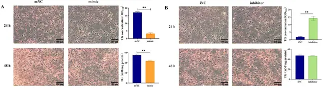
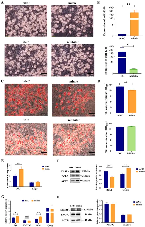
This study aims to investigate the effect of miR-130b on lipid accumulation at early-stage (24 and 48 h) and late-stage (7 d) adipogenic differentiation and to clarify potential mechanisms using primary rat intramuscular preadipocytes (IMAs).
Results showed that at 24 h and 48 h, miR-130b overexpression significantly reduced lipid deposition by inhibiting proliferation and inducing apoptosis. Furthermore, miR-130b overexpression significantly inhibited the expression of cell cycle and apoptosis marker genes. Specifically, the mRNA expression of Ccnd1 tended to decrease, while the BCL2 protein level was significantly decreased at 48 h. In contrast, miR-130b inhibition significantly increased the BCL2 protein level. At 7 d, the miR-130b mimic significantly decreased intracellular TG content and tended to decrease Hsd11b1 mRNA expression while significantly promoting Lpl mRNA expression. Additionally, the miR-130b mimic significantly increased the CASP3 protein level and tended to decrease the BCL2 protein level.
In conclusion, these data indicated for the first time that miR-130b could reduce lipid deposition in rat IMAs through different mechanisms: at the early stage of differentiation by inhibiting proliferation and promoting apoptosis and at the late stage by inhibiting adipogenic differentiation, promoting lipid hydrolysis, and promoting apoptosis.
Ask a Question
Write your own review
Description: The thoracic aorta is located in the chest cavity and gives off arteries that branch to the esophagus, pericardium, lungs, and trachea. The thoracic aorta can be subdivided into the ascending aorta, ...
Description: Rat Podocytes are isolated from normal rat kidney. The cells are characterized by immunofluorescence with antibodies specific to podocin, Ang1, Nephrin, ACTN4, NPHS2. T25 flasks is required for cell ...
Description: Rat Bronchial Smooth Muscle Cells are isolated from normal rat bronchi tissue. Rat Bronchial Smooth Muscle Cells are characterized by immunofluorescence with antibodies specific to alpha-actin. T25 ...
Description: Guinea Pig Endothelial Cells from Creative Bioarray are isolated from guinea pig tissue. Prior to shipping, cells at passage 2 are detached from flasks and immediately cryopreserved in vials. Each ...
Description: Rat Lung Epithelial Cells are isolated from normal rat lung tissue. The cells are characterized by immunofluorescence with antibodies specific to CK-18, CK-19. T25 flasks is required for cell ...
Description: Rat Vein Endothelial Cells from Creative Bioarray are isolated from inferior vena cava tissue of 6-8 week old laboratory Sprague-Dawley rat. Rat Vein Endothelial Cells are grown in T75 tissue culture ...